

Summary Card
Overview of Facial Nerve Anatomy
Cranial nerve VII is a motor and sensory nerve with intracranial, intratemporal, and extratemporal anatomical sections.
Intracranial Facial Nerve Anatomy
Originates at the medullopontine angle of the brain stem from the facial nerve proper and nervus intermedius.
Intratemporal Facial Nerve Anatomy
The intratemporal facial nerve enters the internal auditory canal and provides motor, sensory, and secretomotor branches.
Extracranial Facial Nerve Anatomy
Begins at the stylomastoid foramen, provides branches to nearby muscles, enters the parotid, and divides into the 5 terminal branches.
Terminal Branches of the Facial Nerve
Mnemonic: To Zanzibar By Motor Car: Temporal, Zygomatic, Buccal, Marginal Mandibular, Cervical.
Overview of Facial Nerve Anatomy
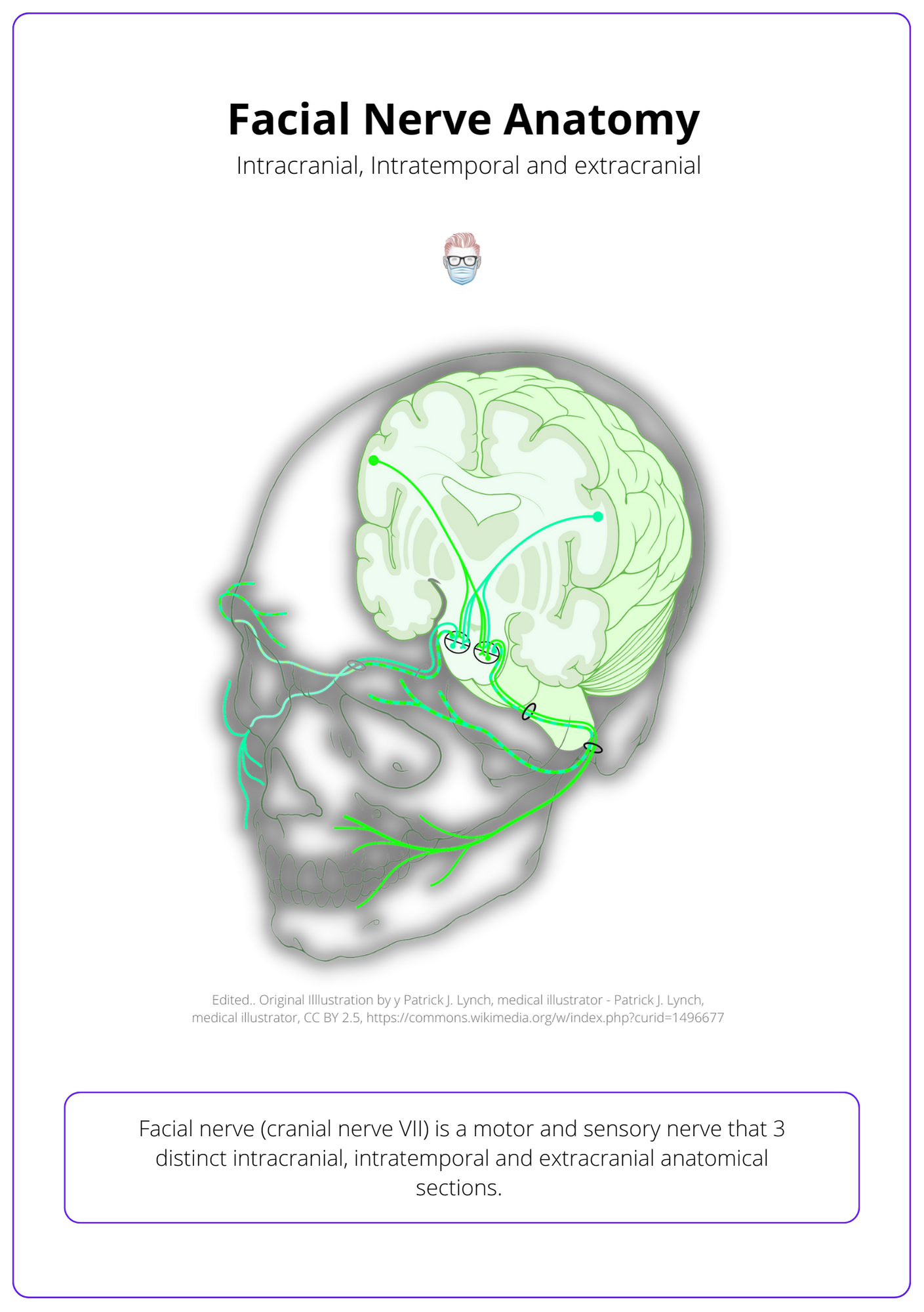
The facial nerve (cranial nerve VII) is a motor and sensory nerve that has 3 distinct intracranial, intratemporal, and extratemporal anatomical sections.
The facial nerve (cranial nerve VII) is a motor and sensory nerve that has 3 distinct intracranial, intratemporal, and extratemporal anatomical sections.
- Intracranial: arises from the precentral gyrus & travels via dorsolateral pons.
- Intratemporal: fallopian canal's labyrinthine, tympanic & mastoid segments.
- Extra-temporal: from stylomastoid foramen to 5 branches via the parotid.
These three sections of the facial nerve are visualised in the illustration below.
Intracranial Facial Nerve Anatomy
The facial nerve arises from the medullopontine angle of the brain stem from the facial nerve proper (motor) and nervus intermedius (parasympathetic and sensory fibres).
Origin
The facial nerve is formed by the facial nerve proper and the nervous intermedius.
- Facial Nerve Proper: motor fibres exiting from the cerebellopontine angle.
- Nervus Intermedius: parasympathetic and sensory fibres.
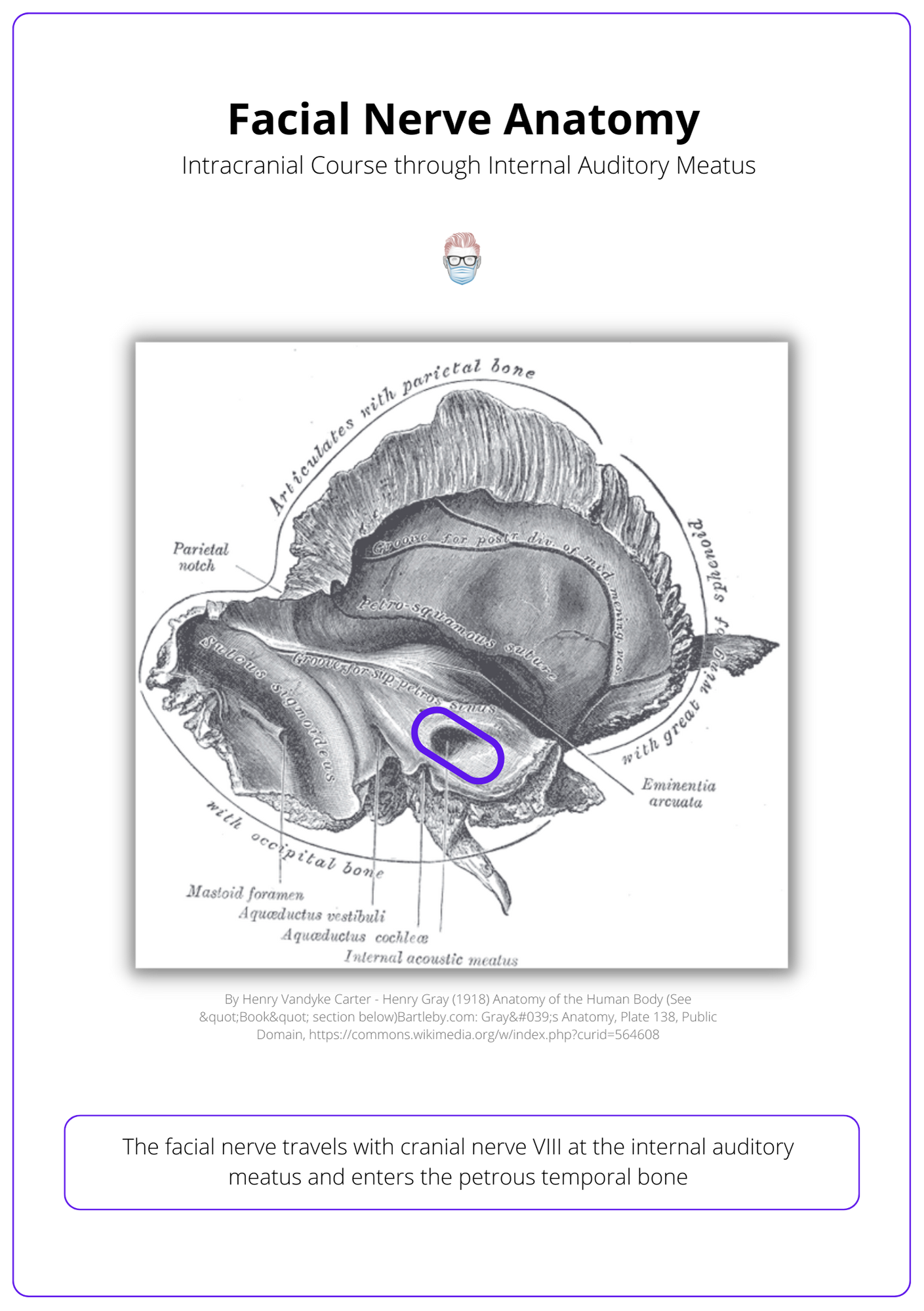
Intracranial Course
The intracranial section has 3 important anatomical landmarks:
- Arises from the medullopontine angle of the brain stem.
- Travels with cranial nerve VIII at the internal auditory meatus.
- Enters the petrous temporal bone.
This intracranial course of the facial nerve is visualised in the illustration below.
Intratemporal Facial Nerve Anatomy
The intratemporal facial nerve enters the internal auditory canal, has 5 main segments, and provides motor, sensory, and secretomotor branches.
Intratemporal Course
- Enters the internal auditory canal with the acoustic and vestibular nerves.
- Travels the fallopian canal by itself through the segments.
- Segments: cisternal, intracanlicular, labyrinthine, tympanic, mastoid.
Intratemporal Branches
Three clinically significant branches from this segment are:
Greater Petrosal:
- Innervates lacrimal gland & soft palate taste via geniculate ganglion.
- Arises in the tympanic segment.
Nerve to Stapedius:
- Dampens loud noises (unaffected in Möbius Syndrome).
- Arises from the mastoid segment.
Chorda Tympani:
- Innervates salivary glands, anterior 2/3 tongue taste via lingual.
- Arises from the mastoid segment.
Extracranial Facial Nerve Anatomy
The extracranial facial nerve arises from the stylomastoid foramen, provides branches to nearby muscles, enters the parotid, and divides into the 5 terminal branches.
Extracranial Course
The facial nerve becomes extracranial after the stylomastoid foramen, where it is protected by the mastoid tip, tympanic ring, and mandibular ramus.
It then travels past bone, muscle, and cartilage before reaching the parotid gland.
- Anterior to the posterior belly of the digastric muscle.
- Lateral to the styloid process of the temporal bone.
- Inferomedially (~1cm3) to the tip of the tragal pointer of the ear.
- Posteriomedially enters the parotid gland.
- Forms temprozygomatic and cervicofacial trunks in the pes anserinus.
- Divides into terminal branches that innervate facial muscles.
Extracranial Branches
The facial nerve provides motor branches to the structures it passes by on its way to the parotid gland.
- Postauricular nerve: occipitalis and posterior auricular muscles
- Branch to the posterior belly of digastric and stylohyoid
- Terminal branches: this is discussed in more detail below.
Terminal Branches of the Facial Nerve
Facial nerve branches can be remembered by the mnemonic: To Zanzibar By Motor Car: Temporal, Zygomatic, Buccal, Marginal Mandibular, Cervical.
Terminal Branches of Facial Nerve
Facial nerve branches can be remembered by the mnemonic: To Zanzibar By Motor Car: Temporal, Zygomatic, Buccal, Marginal Mandibular, Cervical.
- Temporal: innervates the frontalis muscle.
- Zygomatic: innervates the orbicularis oculi and upper lip elevators.
- Buccal: innervates buccinator and upper lip muscles.
- Marginal: innervates lower lip mouths.
- Cervical branch: platysma.
Course of Branches
The terminal branches of the facial nerve exit the parotid gland deep into the subcutaneous musculoaponeurotic system (SMAS) layer.
Some branches of the facial nerve have vital landmarks:
- Temporal nerve: Pitanguy's Line (0.5cm below tragus to 1.5cm above lateral brow) in temporoparietal fascia near the superficial temporal artery.
- Buccal branch: travels with the parotid duct (Stenson's duct), Zucker's point.
- Marginal mandibular: inferior border of the mandible, deep to the platysma and superficial to the facial artery and anterior facial vein.
Variations in Facial Nerve
Six distinctive anastomotic patterns were initially classified by Davis et al. in 1956, but there is significant variation in the facial nerve due to:
- No consistent spatial or topographic orientation.
- Variations in branching patterns and anastomoses between branches (Lineaweaver et al., 1997 & Kwak et al., 2004).
- The temporal branch and marginal mandibular nerve have less arborization than other facial nerve branches.
These branching pattern variations and cross-innervation clinically correlate to unexplained facial muscle strength in the presence of a known injury to the facial nerve.
Conclusion
1. Facial Nerve Anatomy: You have gained comprehensive knowledge of the facial nerve's structure, functions, and three main anatomical sections (intracranial, intratemporal, and extratemporal), along with the variations in its branching patterns.
2. Intracranial Facial Nerve Anatomy: You now understand the intracranial portion, which originates from the medullopontine angle, and includes both the motor facial nerve proper and the sensory/parasympathetic nervus intermedius.
3. Intratemporal Facial Nerve Anatomy: You learned the path and branches of the facial nerve as it travels through the intratemporal segment, supplying motor, sensory, and secretomotor functions through key branches like the greater petrosal, nerve to stapedius, and chorda tympani.
4. Extracranial Facial Nerve Anatomy: You explored the course and branches of the nerve after emerging from the stylomastoid foramen, including the five terminal branches (temporal, zygomatic, buccal, marginal mandibular, and cervical) that control facial muscles.
5. Terminal Branches Mnemonic and Landmarks: You mastered the mnemonic "To Zanzibar By Motor Car" to remember the terminal branches and identify their landmarks to ensure precise clinical identification and intervention.
6. Variations in Branching Patterns: You understand the distinctive anastomotic patterns and variations that influence facial nerve structure, which correlate with facial muscle function even in the presence of nerve injury.
Further Reading
- Greyling L, Glanvill R, Boon J, et al. Bony landmarks as an aid for intraoperative facial nerve identification. Clin Anat. 2007;20(7):739-744. doi:10.1002/ca.20508
- D’Antoni AV. Gray’s Anatomy, the Anatomical Basis of Clinical Practice, Forty-First Edition, by Susan Standring, Editor-in-Chief, Elsevier Limited, 2016, 1,562 Pages, Hardcover, $228.99 ($171.74), ISBN: 978-0-7020-5230-9. Clin Anat. January 2016:264-265. doi:10.1002/ca.22677
- Westphal M. Cranial Neuroimaging and Clinical Neuroanatomy, Third Ed. Neuro-Oncology. July 2004:267-268. doi:10.1215/s1152851704200035
- DAVIS R, ANSON B, BUDINGER J, KURTH L. Surgical anatomy of the facial nerve and parotid gland based upon a study of 350 cervicofacial halves. Surg Gynecol Obstet. 1956;102(4):385-412. https://www.ncbi.nlm.nih.gov/pubmed/13311719.
- Lineaweaver W, Rhoton A, Habal M. Microsurgical anatomy of the facial nerve. J Craniofac Surg. 1997;8(1):6-10. https://www.ncbi.nlm.nih.gov/pubmed/10332290.
- Kwak H, Park H, Youn K, et al. Branching patterns of the facial nerve and its communication with the auriculotemporal nerve. Surg Radiol Anat. 2004;26(6):494-500. doi:10.1007/s00276-004-0259-6
