

Summary Card
Lip Neurovascular Anatomy
A composite soft tissue structure innervated by branches of the facial nerve and trigeminal nerve, supplied by labial arteries.
Anatomical Lip Landmarks
A specific set of landmarks includes the vermillion border, cupid's bow, philtrum, wet-dry mucosa, white roll, tubercle, and commissure.
Aesthetic Units of the Lip
The aesthetic subunits of the lip can be described as upper lateral, philtrum, lower lip, chin, and cheek.
Muscles of the Lip
Act as depressors & elevators. Most insert into the modiolus and are innervated by buccal & mandibular branches of the facial nerve.
Lip Neurovascular Anatomy
The lip is a composite soft tissue structure that receives motor innervation from branches of the facial nerve and sensation from trigeminal nerve branches.
The lip is a structure composed of skin, fat, muscle, and mucosa. The lip plays a vital role in oral competence, sensation, speech, and cosmesis.
The neurovascular anatomy of the lip is detailed below.
- Motor: buccal and mandibular branches of the facial nerve.
- Sensory: infraorbital (upper) & mental (lower) branches of trigeminal nerve.
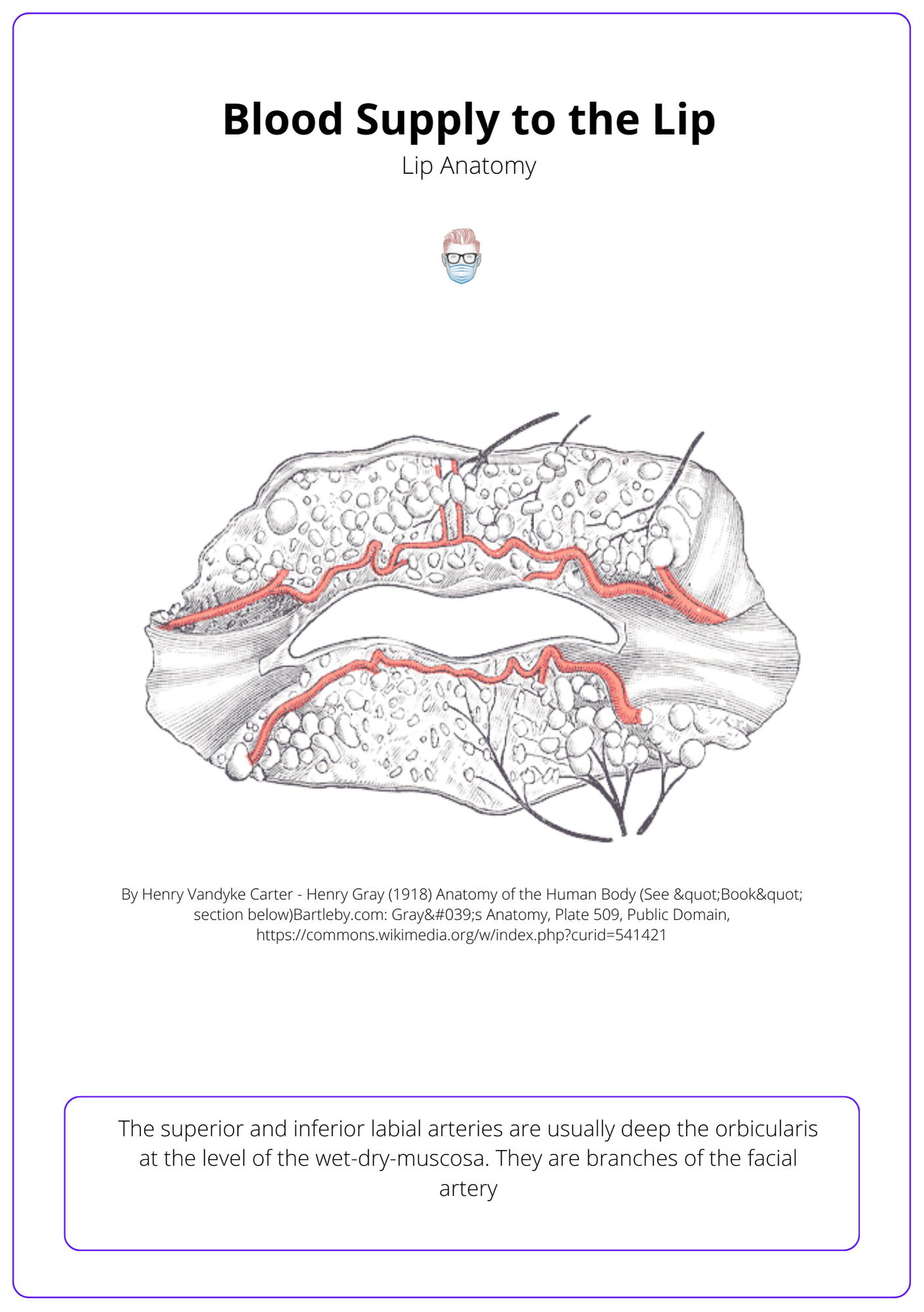
- Blood supply: superior and inferior labial branches of the facial artery.
- Lymph: ipsilateral submandibular nodes; the middle can drain to submental.
The image below illustrates the blood supply to the lip.
Anatomical Lip Landmarks
The lip has specific anatomical landmarks that should be respected during the reconstructive process. This includes the vermillion border, cupid's bow, philtrum, wet-dry mucosa, white roll, tubercle, and commissure.
During the reconstruction of the lip in both elective and non-elective cases, it's important to identify key landmarks. This includes commissure, cupid's bow, tubercle and the red line (wet-dry vermillion line).
These anatomical landmarks of the lip can be visualised below.

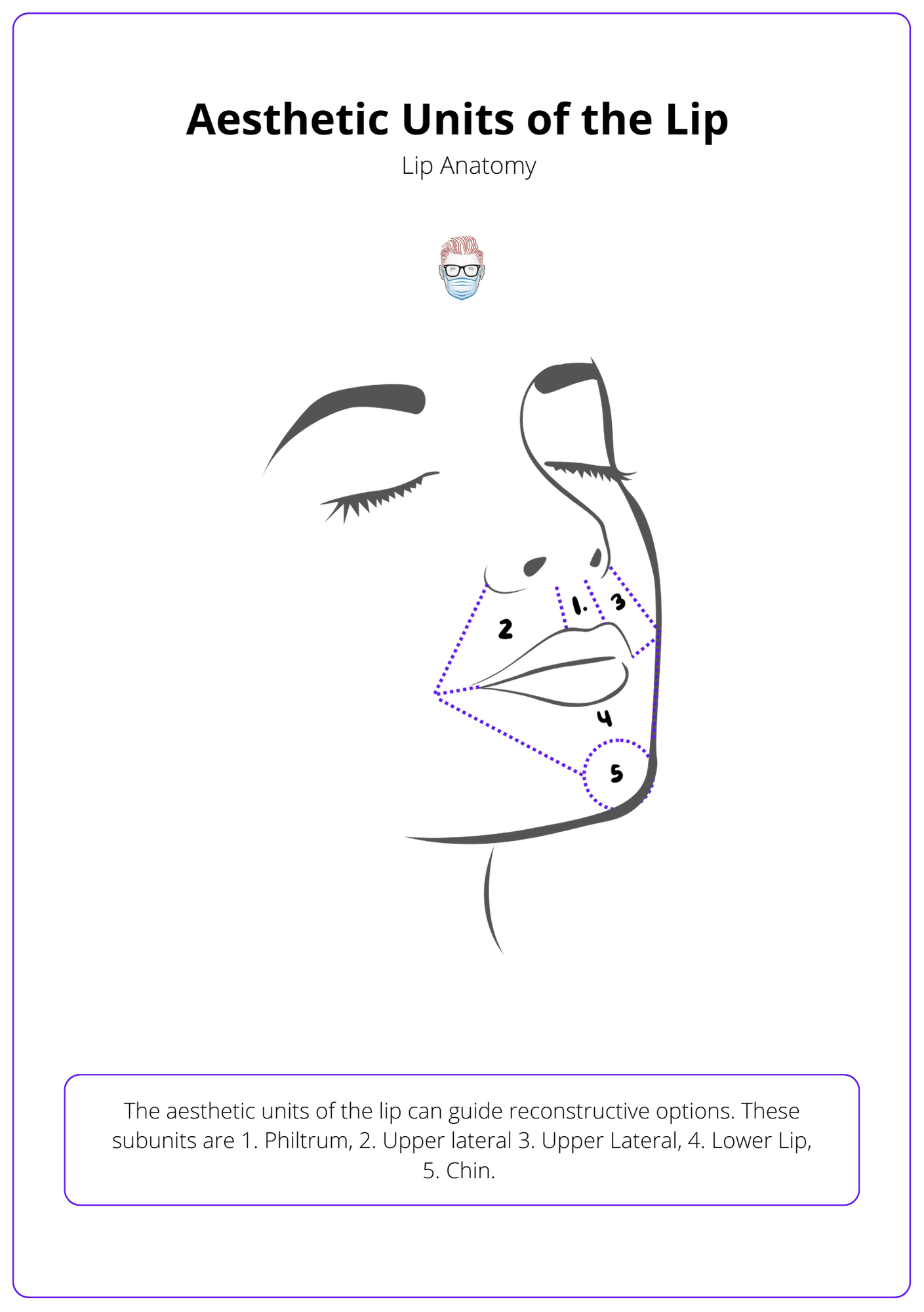
Aesthetic Units of the Lip
The aesthetic subunits of the lip can be described as upper lateral, philtrum, lower lip, chin, and cheek.
The face has several areas of subunits to help with treatment algorithms and reconstructive options – the lip is no different. There are 5 aesthetic units directly relevant to the lip (six if you include the cheek).
These aesthetic subunits can be visualised in the diagram below.
Muscles of the Lip
The muscles of the lip act as depressors and elevators. The majority of the muscles insert into the modiolus and are innervated by the buccal and mandibular branches of the facial nerve.
The muscles of the lip can be classified into elevators and depressors. Their primary innervation is through the buccal and mandibular branches. These muscles may be damaged during trauma, localised for botox or reconstructed in dynamic facial nerve surgeries.
The image below illustrates the facial expression muscles.
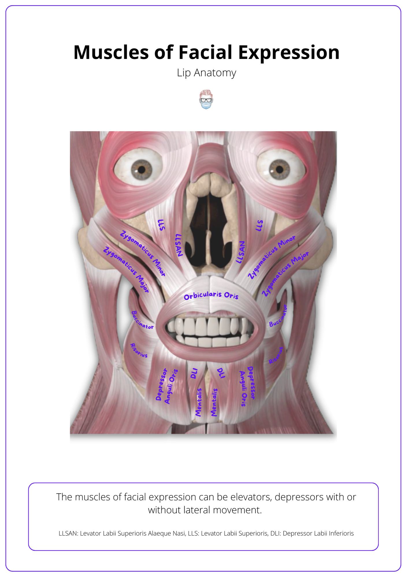
These muscles have specific origins and insertion points on the face. This clues the type of function (elevation or depression) and nerve innervation (buccal or mandibular branch).
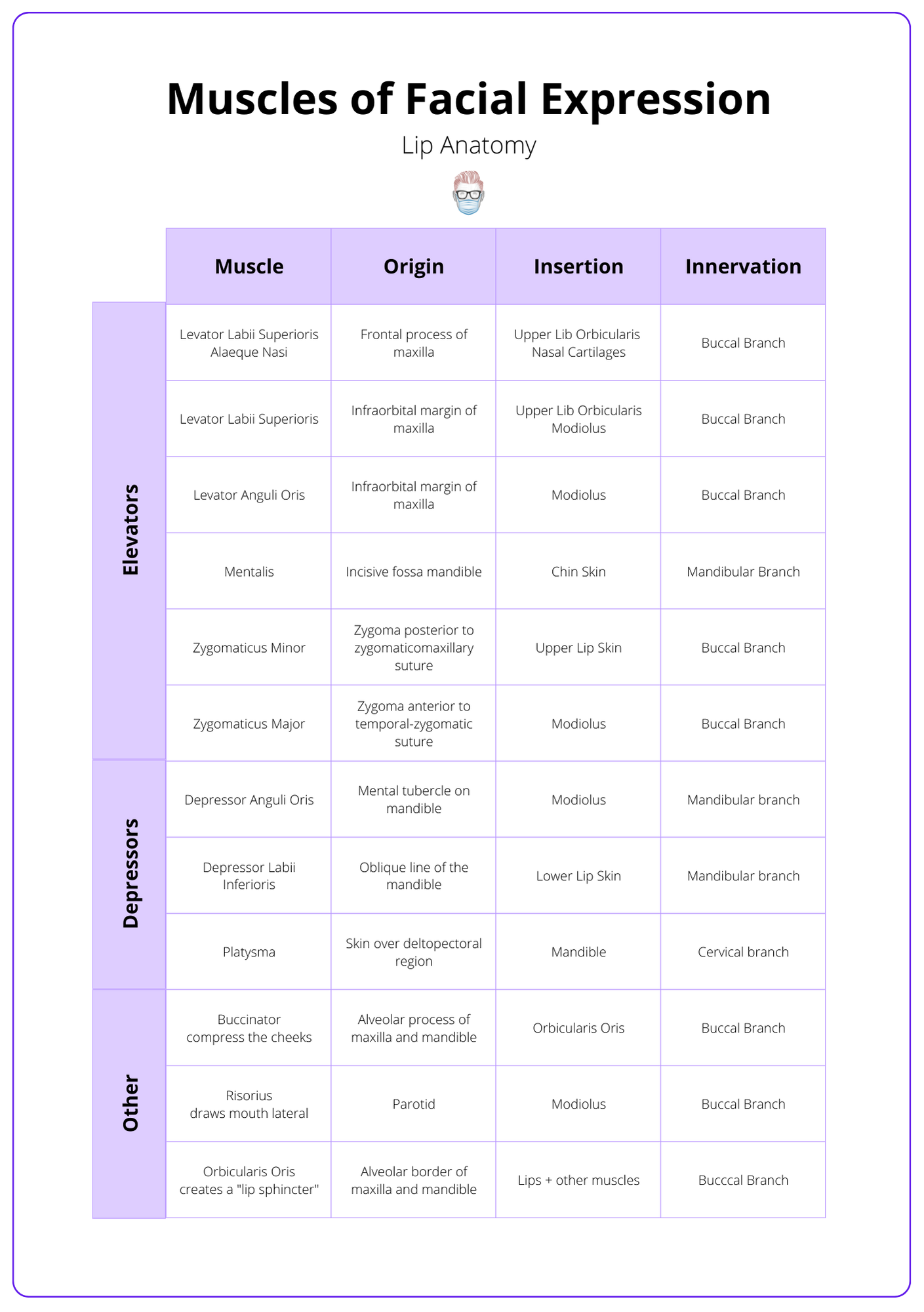
These lip muscle origin and insertion points can be seen in the table below.
Conclusion
1. Lip Neurovascular Anatomy: You have a comprehensive grasp of the lip's neurovascular anatomy, including the motor innervation from the facial nerve branches, sensory input from trigeminal branches, and blood supply from the labial arteries.
2. Anatomical Landmarks: You know the key anatomical landmarks of the lip, such as the vermillion border, cupid's bow, philtrum, wet-dry mucosa, white roll, tubercle, and commissure, essential for reconstructive and aesthetic procedures.
3. Aesthetic Units: You understand the aesthetic units of the lip, including upper lateral, philtrum, lower lip, chin, and cheek, to guide reconstructive and cosmetic planning.
4. Muscle Function and Innervation: You have detailed knowledge of the muscles of the lip, their role as elevators and depressors, and their innervation through the buccal and mandibular branches.
5. Clinical Tips and Techniques: You have acquired practical tips on the anatomical structures and their importance in cosmetic and reconstructive surgery, like the significance of the philtrum and modiolus for facial muscle function.
