

Summary Card
Cervical Lymphatic Drainage Pattern
The lymphatic flow is from superficial to deeper and from the upper to lower parts of the neck.
Level 1 Cervical Lymph Nodes
Superior: Mandible
Inferior: Anterior and posterior digastric muscle, hyoid.
Walls: N/A
Structures: Submental nodes, submandibular nodes & glands.
Level 2 Cervical Lymph Nodes
Superior: Jugular fossa
Inferior: Hyoid bone
Walls: Sternohyoid to sternocleidomastoid
Structures: Spinal accessory nerve divides 2A & 2B
Level 3 Cervical Lymph Nodes
Superior: Hyoid bone
Inferior: Lower border of the cricoid
Walls: Sternohyoid to sternocleidomastoid
Structures: N/A
Level 4 Cervical Lymph Nodes
Superior: Lower border of the cricoid
Inferior: Clavicle
Walls: Sternohyoid to sternocleidomastoid
Structures: Thoracic duct on the left
Level 5 Cervical Lymph Nodes
Superior: Cranium at convergence of SCM & trapezius
Inferior: Clavicle
Walls: Posterior border of SCM and anterior border of trapezius.
Key Contents: Spinal accessory nerve.
Level 6 Cervical Lymph Nodes
Superior: Hyoid bone
Inferior: Suprasternal notch
Walls: Carotid sheaths
Structures: Pre- and para-tracheal nodes.
Level 7 Cervical Lymph Nodes
Superior: Suprasternal notch
Inferior: Aortic arch
Walls: Carotid sheaths
Sublevel: None
Cervical Lymphatic Drainage Pattern
The lymphatic flow is from superficial to deeper and from the upper to lower parts of the neck. The lymphatic system of the neck is divided into three systems.
Overview
In general, the lymphatic flow is from superficial to deeper and from the upper to lower parts of the neck. The lymphatic system of the neck is divided into three systems:
- Waldeyer’s internal ring: Nodes in adenoids, tonsils, and posterior pharyngeal wall which are at high risk for lymphatic spread.
- Waldeyer’s external ring: Nodes in the occipital, post-auricular, parotid, superficial cervical, submandibular, submental, and anterior cervical region. They drain superficial tissues from the scalp, eyelids, face, sinuses, and oral cavity.
- Deep lymph node system: Nodes in the upper, middle, and lower cervical regions near the internal jugular vein, a spinal accessory group, and neck midline.
Drainage Patterns
Each anatomical subgroup of lymph nodes acts as a reservoir for a specific site in the head and neck region. This creates the potential to identify the primary tumour based on the site of a specific group of lymph nodes.
- Level 1: Lips, gums, teeth, tongue, and anterior hard palate
- Level 2: Naso-/oro-/hypopharynx and parotid
- Level 3: Naso-/oro-/hypopharynx and larynx
- Level 4: Larynx, cervical oesophagus, and hypopharynx
- Level 5: Naso- and oropharynx
- Level 6: Thyroid, parathyroid, cervical oesophagus, and larynx
Clinically speaking, there are some generally accepted pathological draining routes. For example:
- The floor of mouth and tongue tumors tend to spread to levels I through III
- Laryngeal tumors tend to spread to levels II through IV
Level 1 Cervical Lymph Nodes
Level 1 cervical lymph nodes contain the submental (1A) and submandibular (1B) triangles.
Level 1 cervical lymph nodes have two sublevels: submental (1A) & submandibular (1B). This can be visualized in the illustration below.

Level 1A: Submental
- Location: Between anterior digastric bellies and hyoid bone
- Structures: Submental nodes
- Drainage: From lips, gum, tongue, anterior hard palate
Level 1B: Submandibular
- Location: Between the digastric muscle and the body of the mandible
- Structures: Submandibular nodes and gland
- Drainage: From lips, gum, tongue, anterior hard palate
Level 2 Cervical Lymph Nodes
Upper jugular neck nodes have anterior (2A) & posterior (2B) divisions.
Upper jugular neck nodes have anterior (2A) & posterior (2B) divisions. This is illustrated in the image below.

The location of level 2 cervical neck nodes are:
- Superior: Skull base at jugular fossa
- Inferior: Hyoid bone or carotid bifurcation
- Posterior: Posterior border sternocleidomastoid (SCM)
- Anterior: Continuation from level 1B (lateral border of the sternohyoid or posterior belly of digastric depending on resource)
Key points on level 2 neck nodes are:
- Contents: Spinal accessory nerve and upper jugular nodes
- Divisions: Anterior (2A) & posterior (2B) divided by spinal accessory nerve
- Drainage: Naso-/oro-/hypopharynx and parotid
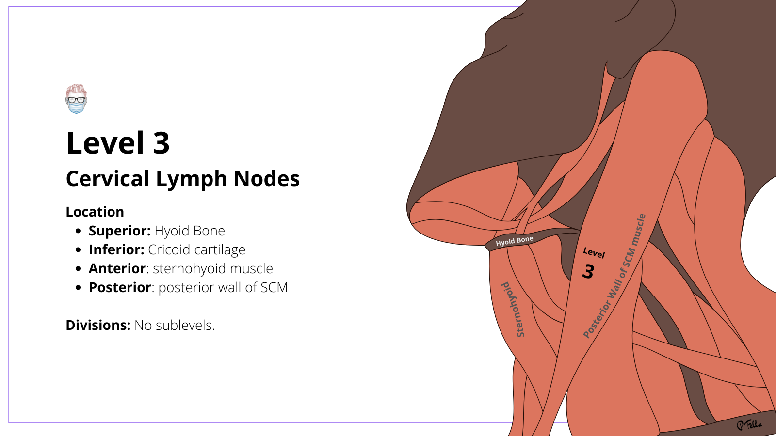
Level 3 Cervical Lymph Nodes
The middle jugular group does not have sublevels.
The middle jugular group does not have sublevels. The location of level 3 cervical nodes are:
- Superior: Hyoid bone or carotid bifurcation
- Inferior: Lower cricoid or superior belly of the omohyoid muscle
- Anterior: Sternohyoid (don't get confused with stylohyoid)
- Posterior: Posterior border of sternocleidomastoid (SCM)
Level 3 tends to drain the naso-/oro-/hypopharynx and larynx.
Level 4 Cervical Lymph Nodes
The lower jugular group has 4A & 4B sublevels containing the thoracic duct and Virchow's node.
The lower jugular group has 4A & 4B sublevels containing the thoracic duct and Virchow's node. This is illustrated in the image below.

The location of level 4 cervical nodes are:
- Superior: Cricothyroid notch or omohyoid muscle
- Inferior: Clavicle
- Anterior: Lateral border of sternohyoid muscle (same as level 3)
- Posterior: Posterior border of sternocleidomastoid (same as level 3)
The sublevels are described as follows:
- Divisions: 4A is deep to sternal SCM, 4B is deep to clavicular SCM
- Contents: Lower jugular nodes, left thoracic duct, right lymphatic duct
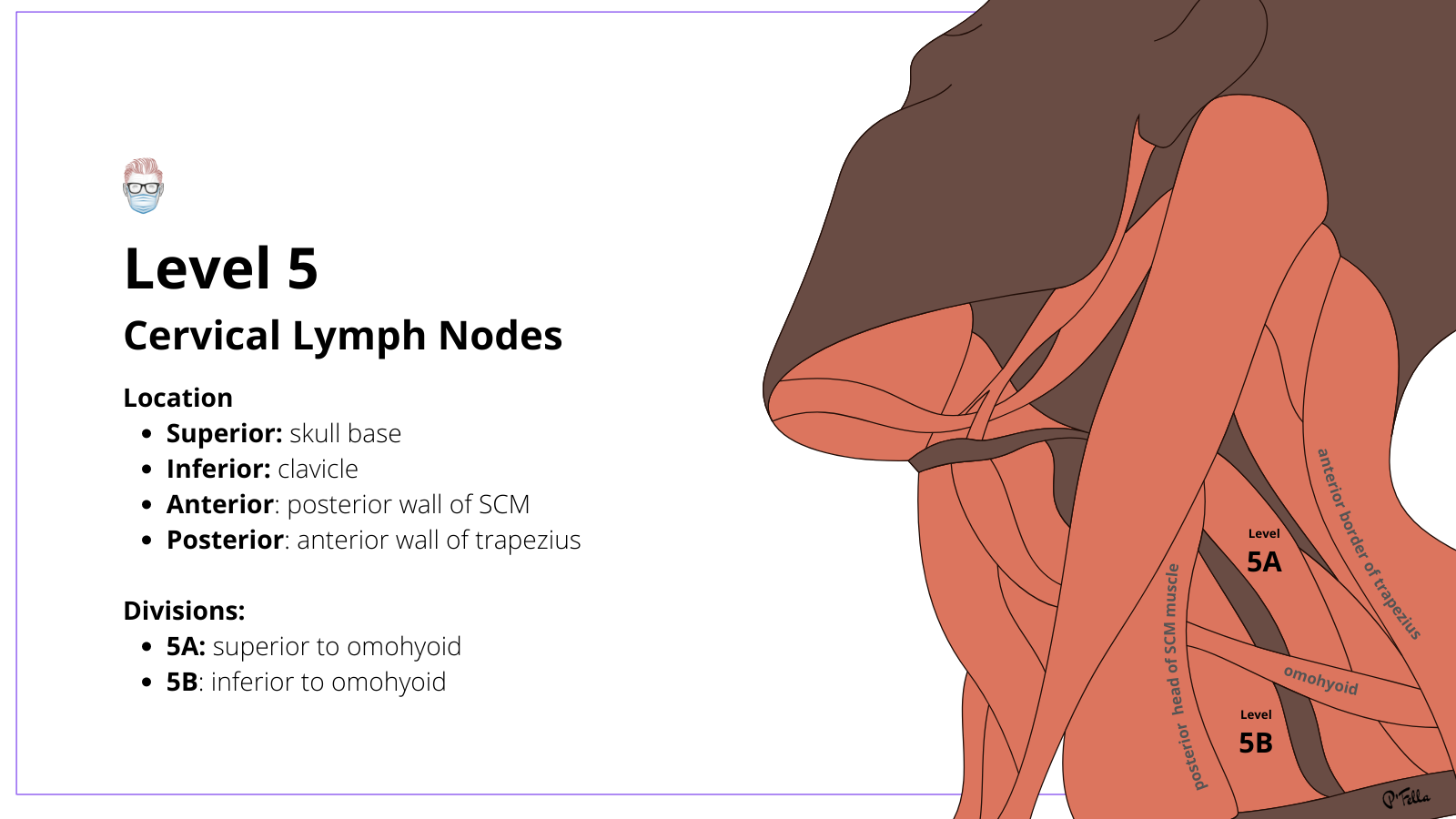
Level 5 Cervical Lymph Nodes
The posterior triangle group is divided into sublevels 5A & 5B by omohyoid.
The posterior triangle group is divided into sublevels 5A & 5B by omohyoid. Level 5 cervical neck lymph nodes drain the naso- and oro-pharynx.
The anatomical locations are:
- Superior: Skull base SCM and trapezius muscles
- Inferior: Clavicle
- Anterior: Posterior border of the SCM muscle
- Posterior: Anterior border of the trapezius muscle
The sublevels are described as follows:
- 5A: Superior to the omohyoid and contains the spinal accessory nodes
- 5B: Inferior to the omohyoid and contains the transverse cervical and supraclavicular nodes
Level 6 Cervical Lymph Nodes
The anterior compartment contains the paratracheal and Delphian nodes.
The anterior compartment contains the paratracheal and Delphian nodes. Level 6 is medial to levels III and IV. It drains the thyroid, parathyroid, cervical oesophagus, and larynx. The anatomical landmarks are:
- Superior: Hyoid bone
- Inferior: Manubrium/suprasternal notch
- Walls: Left and right common carotid arteries

Level 7 Cervical Lymph Nodes
Upper mediastinal nodes lie outside the borders of the neck.
Upper mediastinal nodes lie outside the borders of the neck. The anatomical location of Level VII nodes are:
- Superior: Suprasternal notch
- Inferior: Aortic arch
- Lateral: Carotid arteries
These nodes are an extension of the paratracheal lymph node chain.
Conclusion
1. Basics of Cervical Lymph Nodes: Gained an understanding of the anatomical arrangement of cervical lymph nodes and their significance in lymphatic drainage in the neck region.
2. Anatomical Landmarks: Learned about the anatomical landmarks that define each cervical lymph node level.
3. Lymphatic Drainage Patterns: Recognized the specific drainage patterns associated with each group of lymph nodes, crucial for diagnosing the primary sites of tumors.
4. Clinical Implications: Understood the clinical implications of lymphatic drainage in the management of various head and neck conditions.
5. Surgical Relevance: Reviewed the importance of these lymph nodes in surgical planning, particularly in oncology, to ensure comprehensive management of malignancies in the head and neck.
References
- Haynes J, Arnold K, Aguirre-Oskins C, Chandra S. Evaluation of neck masses in adults. Am Fam Physician. 2015;91(10):698-706.
- Robbins K, Shaha A, Medina J, et al. Consensus statement on the classification and terminology of neck dissection. Arch Otolaryngol Head Neck Surg. 2008;134(5):536-538. doi:10.1001/archotol.134.5.536
- Lymph nodes of the neck. P M SomRadiology1987165:3, 593-600
- Martin G. Mack, Jörg Rieger, Mehran Baghi, Sotirios Bisdas, Thomas J. Vogl,
Cervical lymph nodes, European Journal of Radiology, Volume 66, Issue 3,
2008, Pages 493-500, ISSN 0720-048X, https://doi.org/10.1016/j.ejrad.2008.01.019. - Farr HW, Goldfarb PM, Farr CM. Epidermoid carcinoma of the mouth and pharynx at Memorial Sloan-Kettering Cancer Center, 1965 to 1969. Am J Surg. 1980 Oct;140(4):563-7. doi: 10.1016/0002-9610(80)90213-5. PMID: 7425241.
