


Genioplasty is considered for aesthetic chin imbalance, functional chin-related problems, adjunctive profile correction, and selected airway indications; modify or defer treatment when occlusion, skeletal maturity, disease activity, dental health, or expectations make isolated chin surgery unsafe or misleading.
Applied Anatomy
Safe genioplasty depends on the mandibular symphysis, mental foramina and nerve loops, tooth apices, inferior border, mentalis muscle, labiomental fold, and soft-tissue pogonion.
Assessment and Planning
Assess facial proportions, occlusion, cephalometry, 3D bony anatomy, chin projection, vertical height, asymmetry, soft-tissue thickness, airway context, photographs, and patient goals before choosing the vector or device.
Procedure Selection
Choose implants or fillers for volume-dominant mild deficiency, osseous genioplasty for vector-based skeletal change, reduction or vertical movement for dimensional excess, asymmetric osteotomy for true bony asymmetry, and orthognathic surgery when occlusion or jaw relationship is the primary deformity.
Operative Principles
Safe genioplasty requires accurate marking, intraoral exposure, mental nerve protection, root-safe osteotomy design, controlled three-dimensional segment movement, secure fixation, selective grafting, mentalis resuspension, and final symmetry checks.
Complications
Complications include bleeding, infection, dehiscence, neurosensory disturbance, dental injury, malposition, relapse, nonunion, implant failure, soft-tissue ptosis, asymmetry, and revision for exposed, infected, unstable, or aesthetically unacceptable results.
Postoperative Care and Surveillance
Monitor airway, bleeding, oral wound integrity, fixation or implant position, sensation, occlusion, and infection; escalate dyspnoea, expanding swelling, fever, wound breakdown, exposure, malocclusion, or progressive numbness.
Outcomes and Counselling
Genioplasty can improve lower facial balance and selected functional goals, but soft-tissue prediction is imperfect, osseous results are generally durable, relapse and revision remain possible, and implants need device-lifecycle counselling.
Indications and Suitability
Genioplasty is considered for aesthetic chin imbalance, functional chin-related problems, adjunctive profile correction, and selected airway indications; modify or defer treatment when occlusion, skeletal maturity, disease activity, dental health, or expectations make isolated chin surgery unsafe or misleading.
Genioplasty changes the chin point, lower facial contour, and soft-tissue support. The first decision is whether the chin is truly the problem, or whether it is a visible marker of a larger dentofacial deformity.
Patient Selection
Offer genioplasty when the chin deformity is anatomically correctable and the planned change matches the patient’s functional or aesthetic goal.
- Aesthetic imbalance: chin retrusion, excess, asymmetry, vertical disproportion, or contour deficiency may justify surgery 1.
- Adjunctive profile surgery: genioplasty may refine facial profile in adults undergoing broader aesthetic or orthognathic planning 2.
- Airway context: genioplasty can be selected as a functional procedure in obstructive sleep apnoea reconstruction pathways 3.
- Not rhinoplasty-only: chin correction may be indicated for facial balance beyond rhinoplasty planning 4.
Suitability Boundaries
Choose the operation only after deciding whether the required correction is soft-tissue volume, bony movement, or occlusal skeletal correction.
- Mild deficiency: implant augmentation may suit mild-to-moderate deficiency when the patient prioritises lower invasiveness, faster recovery, and less discomfort 5.
- Three-dimensional deformity: osseous genioplasty is better suited to versatile movement and complex 3D chin deformity 5.
- Airway goal: osseous advancement can enlarge the airway-related skeletal framework, while an implant cannot advance the genial tubercle 5.
- Contraindications: defer for active oral infection, unstable periodontal disease, unrealistic expectations, or untreated dental pathology.
Occlusal Considerations
Do not use isolated genioplasty to camouflage a malocclusion that needs orthognathic correction.
- Class II pattern: assess mandibular deficiency, overjet, and airway symptoms before offering chin-only augmentation.
- Class III pattern: avoid setback genioplasty as camouflage if maxillomandibular correction is required.
- Skeletal maturity: elective skeletal correction is usually delayed until growth stability is clear.
- Inflammatory disease: in juvenile idiopathic arthritis deformity, orthognathic correction should be considered only when skeletally mature with controlled or quiescent disease and stable deformity 6.
Applied Anatomy
Safe genioplasty depends on the mandibular symphysis, mental foramina and nerve loops, tooth apices, inferior border, mentalis muscle, labiomental fold, and soft-tissue pogonion.
The chin is a small operative field with little margin for error. A millimetric planning mistake can injure roots, denervate the lower lip, destabilise fixation, or create soft-tissue ptosis.
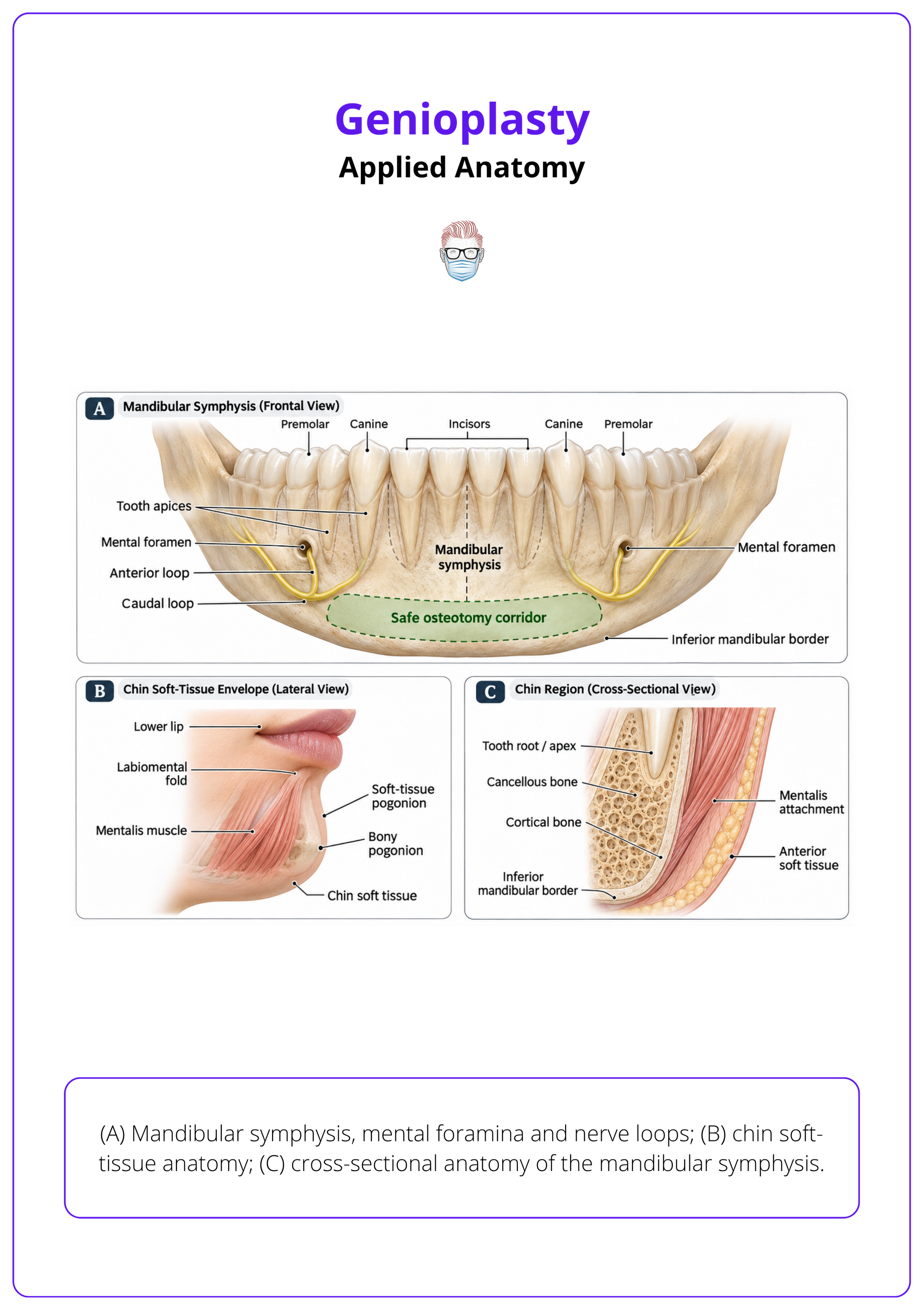
Mandibular Symphysis
Plan the osteotomy inside the symphyseal safe corridor between tooth apices, mental foramina, and the inferior mandibular border.
- Tooth apices: symphyseal planning should respect the vertical distance from tooth apex to mandibular base, reported as 20.3 ± 3.1 mm in one normal-occlusion CBCT sample 7.
- Inferior border: maintain a strong caudal segment for mobilisation, fixation, and contour continuity.
- Cortical thickness: anterior symphyseal cortical thickness was 2.3 ± 0.5 mm in one CBCT sample, relevant to screw purchase 7.
- Step deformity: lateral bony steps occur when the osteotomy or advancement is not feathered into the mandibular body.
Mental Nerve
Preserve the mental nerve by identifying the foramina, anticipating nerve loops, and keeping osteotomy and fixation safely away.
- Foramen position: mental foramina were below the second premolars in 85% of normal-occlusion subjects, with mean interforaminal distance 53.1 ± 3.6 mm 7.
- Anterior loop: operative planning should account for an anterior loop in about 51% of patients, with mean length 2.08 mm 8.
- Caudal loop: the caudal loop should be treated as a consistent landmark, with pooled prevalence 100% and mean length 4.73 mm 8.
- Danger zone: planned osteotomy and hardware should preserve distance from anterior and caudal mental nerve loops 8.
Soft-Tissue Envelope
The final chin result depends on muscle support and soft-tissue drape, not bone position alone.
- Soft pogonion: the visible chin point reflects bony pogonion plus soft-tissue thickness and tone.
- Labiomental fold: deepening, flattening, or asymmetry can follow excessive movement or poor soft-tissue handling.
- Mentalis: mentalis tensioning reduces the risk of chin ptosis and lower lip incompetence 9.
- Sensory risk: mental nerve injury can cause neuropraxia and lower lip or chin sensory deficit 9.
Assessment and Planning
Assess facial proportions, occlusion, cephalometry, 3D bony anatomy, chin projection, vertical height, asymmetry, soft-tissue thickness, airway context, photographs, and patient goals before choosing the vector or device.
Planning is the operation before the operation. Poor planning usually presents later as an apparently “technical” complication: wrong projection, lip incompetence, asymmetry, relapse, or an unhappy patient.
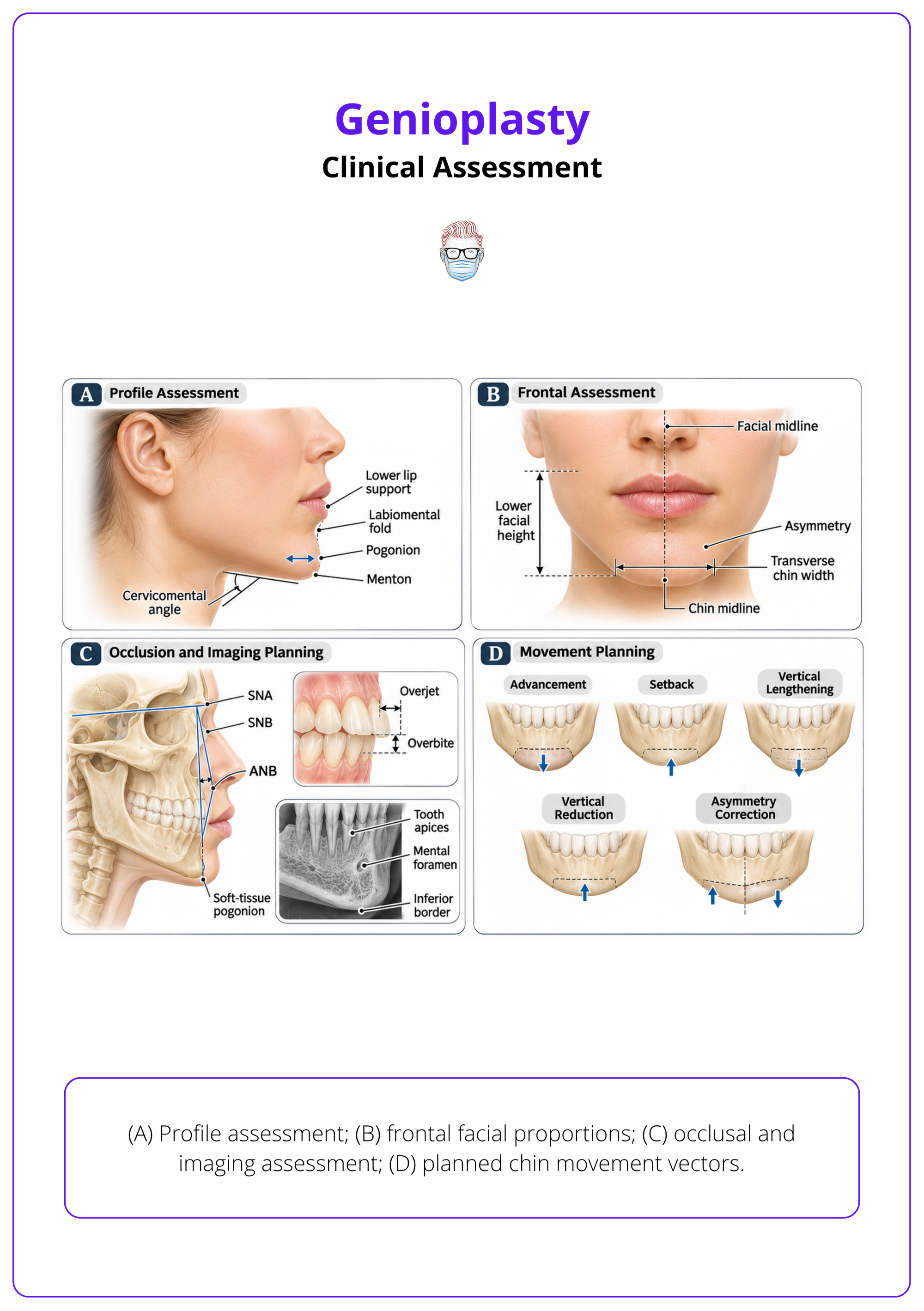
Facial Analysis
Define whether the chin problem is projection, vertical height, transverse width, asymmetry, or soft-tissue contour.
- Profile view: assess pogonion, menton, lower lip support, labiomental fold, and cervicomental angle.
- Frontal view: document chin midline, mandibular cant, lower facial height, and transverse chin width.
- Microgenia grading: cephalometry can classify microgenia using the pogonion-to-zero-meridian horizontal distance 5.
- Severity bands: microgenia may be described as mild when pogonion is retracted <10 mm, moderate at 10–20 mm, and severe at >20 mm 5.
Occlusal Analysis
Treat the chin as part of the dentofacial skeleton, not as an isolated aesthetic island.
- Occlusion: record molar and canine class, overjet, overbite, crossbite, dental midlines, and centric relation discrepancy.
- Orthognathic sequence: when malocclusion is present, genioplasty planning should follow occlusal correction and reassessment of facial morphology 2.
- Maxillary deformity: in dentomaxillary malformation, analyse soft-tissue change after maxillary osteotomy before finalising genioplasty 2.
- Airway history: ask about snoring, witnessed apnoea, CPAP use, prior sleep study, and planned maxillomandibular surgery.
Imaging Planning
Use cephalometry and CBCT to convert aesthetic diagnosis into a safe skeletal movement plan.
- Cephalometry: assess SNA, SNB, ANB, mandibular plane, lower facial height, incisor position, and soft-tissue pogonion.
- CBCT: preoperative CBCT may assist diagnosis, planning, and complication reduction in symphyseal surgery 7.
- Virtual planning: cephalometry, 3D imaging, and virtual surgical planning support selection and operative accuracy 9.
- Safety margins: include the anterior loop, tooth apices, and inferior mandibular border in the osteotomy plan 7.
Soft-Tissue Prediction
Do not promise that soft tissue will move exactly the same distance as bone.
- Advancement: estimate soft-tissue pogonion movement at about 0.9:1 for chin advancement 10.
- Setback: estimate soft-tissue pogonion response at about 0.55:1 for chin setback 10.
- Lengthening: soft pogonion may follow horizontal advancement at 0.9:1 and soft menton vertical extrusion at 0.95:1 10.
- Reduction: setback and vertical reduction show less soft-tissue response than bony movement, around −0.52:1 horizontally and −0.43:1 vertically 10.
Documentation
Record the baseline well enough that the postoperative result can be judged fairly.
- Photography: standardise frontal, oblique, profile, basal, smiling, and animation views.
- Dental record: document occlusion, dental health, orthodontic status, and any restorative risk.
- Patient priorities: separate “stronger chin,” “shorter face,” “less double chin,” and “better profile.”
- Planning caveat: long-term hard-to-soft tissue response is not reliably 1:1, so simulation must be framed as guidance rather than a guarantee 1.
Procedure Selection
Choose implants or fillers for volume-dominant mild deficiency, osseous genioplasty for vector-based skeletal change, reduction or vertical movement for dimensional excess, asymmetric osteotomy for true bony asymmetry, and orthognathic surgery when occlusion or jaw relationship is the primary deformity.
The best procedure is the one that corrects the dominant anatomical problem with the least misleading trade-off. A chin implant is not a substitute for mandibular advancement, and sliding genioplasty is not a substitute for orthodontic-orthognathic correction.
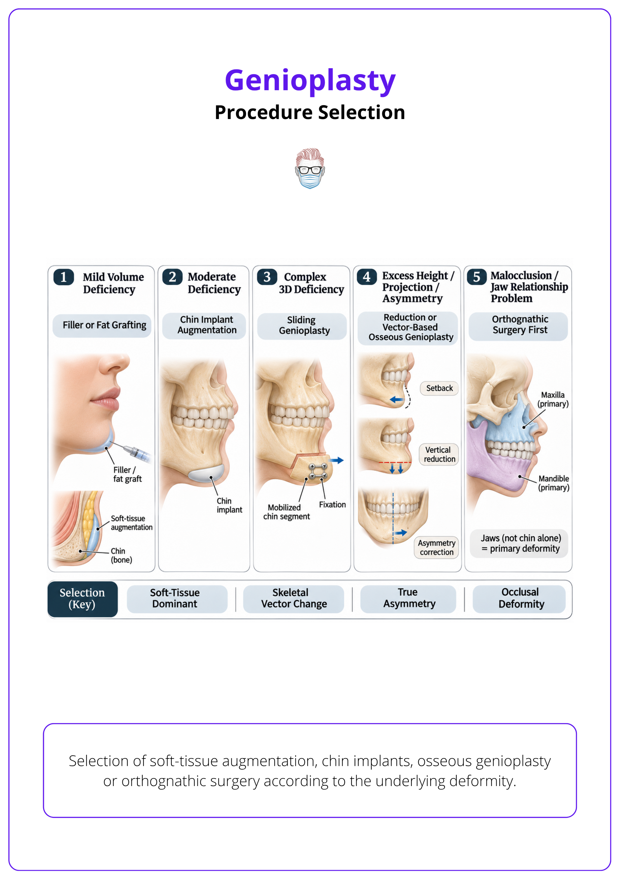
Treatment Selection
Match the intervention to the deformity, durability requirement, and complication profile.
| Dominant problem | Best-fit option | Main trade-off |
|---|---|---|
| Mild volume deficiency | Filler or fat grafting | Limited structural control [c:C46,c:C57] |
| Moderate deficiency | Implant or osseous augmentation | Device risk versus osteotomy morbidity |
| Complex 3D deficiency | Sliding genioplasty | More invasive, more planning-dependent |
| Transverse narrowing or widening | Osseous transverse modification | Soft-tissue prediction is weak 10 |
| Rare anatomical defect | Custom implant | Device lifecycle and cost 11 |
| Multilevel OSA surgery | Broader skeletal strategy | Usually not genioplasty alone |
- No universal winner: satisfaction outcomes do not support one chin augmentation technique as uniquely superior 13.
- Surgical versus nonsurgical: choose by durability and risk profile rather than assuming superiority 14.
- Option range: augmentation options include implants, osteotomy, autologous grafts, fillers, local tissue rearrangements, or combined approaches 13.
Movement Vectors
Select the vector that corrects the measured deformity rather than simply adding projection.
- Advancement: best for retrusive pogonion with acceptable vertical height and occlusion.
- Setback: consider for true chin excess, but reassess Class III jaw relationship first.
- Vertical change: lengthen short lower face or reduce vertical excess using planned bony movement.
- Asymmetry: correct yaw, cant, or unilateral contour by differential osteotomy movement and contouring.
Adjunctive Procedures
Add adjuncts only when they treat a separate visible problem that genioplasty will not solve.
- Submental fullness: liposuction may be added in double-chin patients with mild-to-moderate chin deficiency 15.
- Profile surgery: genioplasty can be performed alone or with rhinoplasty, facelift, or orthognathic surgery 11.
- Autologous grafting: consider fat for mild-to-moderate soft-tissue deficiency, not for major skeletal vector correction 16.
- Implant-plus-osteotomy: reserve combined strategies for selected contour deficits after skeletal movement planning.
Orthognathic Integration
If the bite is wrong, correct the jaws first and use genioplasty to refine the final chin.
- Class II deformity: mandibular advancement or bimaxillary planning may be needed before chin refinement.
- Class III deformity: mandibular setback, maxillary advancement, or bimaxillary correction may change the apparent chin.
- Vertical maxillary excess: lower facial height and lip competence may improve after maxillary surgery.
- Final profile: reassess chin projection after virtual occlusal correction before committing to the genioplasty vector.
Operative Principles
Safe genioplasty requires accurate marking, intraoral exposure, mental nerve protection, root-safe osteotomy design, controlled three-dimensional segment movement, secure fixation, selective grafting, mentalis resuspension, and final symmetry checks.
Most failures come from three errors: unsafe osteotomy level, uncontrolled segment position, or poor soft-tissue repair. The operation should be performed as a skeletal movement with soft-tissue consequences.
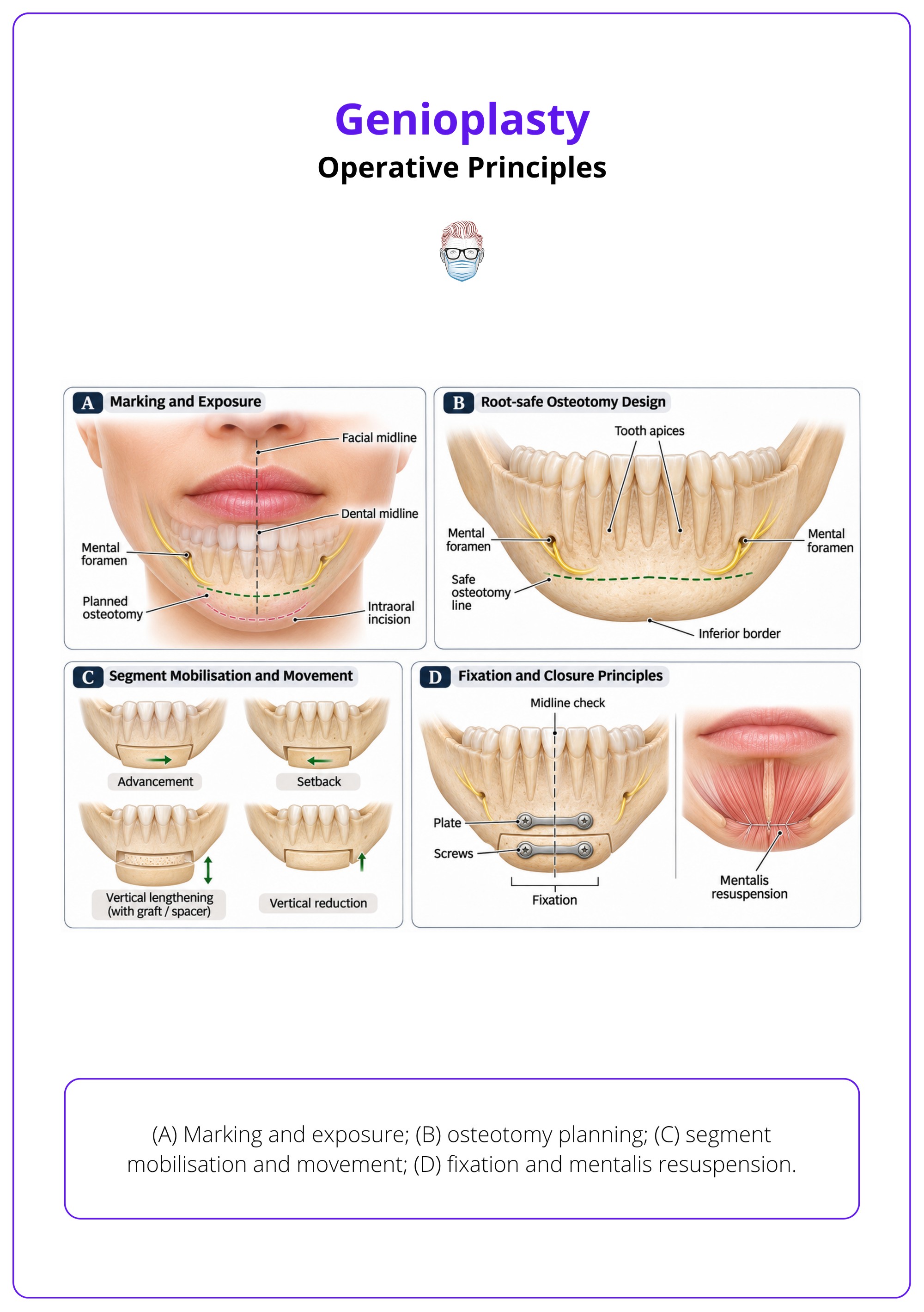
Marking and Exposure
Mark the planned movement and expose enough bone to work safely without stripping the soft-tissue envelope unnecessarily.
- External plan: mark facial midline, dental midline, pogonion, menton, and intended vector.
- Intraoral access: a high labial W-shaped incision may improve access and visibility compared with a curvilinear incision 17.
- Mental nerve: identify the foramina and protect the nerve during lateral exposure and retraction.
- Soft-tissue preservation: limit detachment where possible because periosteal-muscle handling affects predictability 10.
Osteotomy Design
Keep the cut below tooth apices and mental foramina while leaving enough inferior segment for movement and fixation.
- Cut orientation: design the osteotomy to permit the planned advancement, setback, vertical change, or asymmetry correction.
- Three planes: genioplasty can reposition the chin point in all three spatial planes 2.
- Instrument choice: piezotomes may reduce postoperative morbidity and improve long-term satisfaction compared with traditional instruments 18.
- Failure point: an oblique or asymmetric cut can create rotation, step deformity, and midline error.
Segment Mobilisation
Mobilise the segment completely enough to reposition it, but not so aggressively that soft-tissue support is lost.
- Advancement: translate the segment along the planned vector and check midline before fixation.
- Setback or reduction: contour bony edges and avoid excessive soft-tissue redundancy.
- Vertical lengthening: consider interpositional grafting or rigid spacers when bony contact is inadequate.
- Predictability factors: incision, detachment, movement magnitude, fixation, grafting, concomitant jaw surgery, and remodelling all influence soft-tissue response 10.
Fixation and Grafting
Fix the segment rigidly enough to resist muscular recoil and maintain the planned chin point.
- Fixation choice: plates, screws, lag screws, or pre-bent systems are selected according to movement and bony contact.
- Absorbable fixation: resorbable polylactic-polyglycolic acid lag screws or plates can provide satisfactory intraoperative stability 19.
- Rigid fixation: pre-bent absorbable plates or titanium screws are used to improve stability and reduce relapse risk 9.
- Evidence caveat: in isolated advancement genioplasty, reviewed evidence did not link relapse to fixation method or amount of advancement 20.
Closure Checks
Rebuild the soft-tissue envelope before closure and check the result from bone, teeth, lip, and face.
- Mentalis repair: resuspend mentalis securely to reduce ptosis, lip incompetence, and labiomental fold distortion.
- Symmetry check: confirm midline, projection, step-off, dental root safety, and screw position.
- Muscle recoil: secure fixation matters because mandibular soft tissues tend to relapse toward their original position 11.
- Closure: use layered watertight mucosal closure and avoid tension across the intraoral wound.
Complications
Complications include bleeding, infection, dehiscence, neurosensory disturbance, dental injury, malposition, relapse, nonunion, implant failure, soft-tissue ptosis, asymmetry, and revision for exposed, infected, unstable, or aesthetically unacceptable results.
Complications should be separated into urgent threats, structure-specific injuries, and late contour or device problems. Early recognition prevents a small intraoral wound issue becoming hardware exposure or implant loss.
Early Wound Problems
Act early on airway compromise, expanding haematoma, infection, dehiscence, or exposed hardware.
- Bleeding: floor-of-mouth swelling, dysphagia, stridor, or rapidly increasing pain requires urgent review.
- Infection: osseous genioplasty and implants require counselling about infection, hardware or implant problems, bone resorption, and persistent sensory deficits 14.
- Dehiscence: wound breakdown appears more common with implant-based augmentation, reported up to 23.8% versus 5.3% for osseous genioplasty 5.
- Combined surgery: adding genioplasty to Le Fort I and BSSO was not associated with increased length of stay, PONV, opioid use, haematoma, or infection in one cohort 21.
Neurosensory and Dental Injury
Document baseline sensation and investigate persistent, progressive, or anatomically unexpected sensory change.
- Transient nerve symptoms: reviewed chin augmentation complication rates were 15.7% for implants and 19.7% for osteotomy, with transient mental nerve-related injuries in 2.4% and 16.4% respectively 13.
- Persistent deficit: osseous genioplasty has reported transient neurosensory change up to 100% and persistent change in 7.4% to 12.5% 5.
- Dental injury: root injury may present as tooth pain, altered vitality, infection, or radiographic apical damage.
- Neuropathic pain: dysaesthesia, allodynia, or worsening pain needs early senior review and imaging.
Implant and Position Problems
Malposition, asymmetry, relapse, exposed implants, and contour steps require imaging-led diagnosis before revision.
- Plane trade-off: dual-plane alloplastic placement may reduce paraesthesia but has higher malposition, revision, and removal rates than subperiosteal placement 22.
- Access trade-off: intraoral implant access was associated with higher removal rates but lower asymmetry rates than extraoral incisions 22.
- Material trade-off: silicone had lower reported paraesthesia rates than HDPE or ePTFE in reviewed implant literature 22.
- Reoperation: osseous genioplasty reoperation rates ranged from 0% to 2.6%, while implant reoperation rates ranged from 0% to 19% in reviewed studies [c:C49,c:C51].
Postoperative Care and Surveillance
Monitor airway, bleeding, oral wound integrity, fixation or implant position, sensation, occlusion, and infection; escalate dyspnoea, expanding swelling, fever, wound breakdown, exposure, malocclusion, or progressive numbness.
Postoperative care is directed at the intraoral wound, the skeletal construct, and the mental nerves. Patients should understand that swelling and sensory disturbance may obscure the final contour early.
Early Protection
Protect the airway, wound, and fixation during the early inflammatory phase.
- Airway checks: assess voice, swallow, floor-of-mouth swelling, oxygenation, and escalating neck or oral swelling.
- Oral hygiene: use chlorhexidine or local protocol mouth care and avoid traumatic brushing over the incision.
- Diet: use a soft diet initially to reduce wound tension and discomfort during early healing.
- Medication: antibiotics and steroids follow local protocol, implant use, contamination risk, and surgeon preference.
Follow-Up Surveillance
Follow sensation, wound healing, occlusion, and construct position until the result is clinically stable.
- Sensory charting: record lower lip and chin sensation at each visit using side-to-side comparison.
- Recovery adjuncts: L-PRF plus photobiomodulation may improve postoperative mental nerve sensory recovery on two-point discrimination and brush testing 23.
- Expected monitoring: lower lip hypoesthesia should be followed over time; resolution by 6 months was reported in both treated and control sides in one trial 24.
- Implant follow-up: surveillance after chin implant surgery matters because published follow-up is often deficient and complication-rate reliability is uncertain 25.
Outcomes and Counselling
Genioplasty can improve lower facial balance and selected functional goals, but soft-tissue prediction is imperfect, osseous results are generally durable, relapse and revision remain possible, and implants need device-lifecycle counselling.
Counselling should be honest about what the operation changes: chin point, contour, labiomental relationship, and cervicomental balance. It should also be clear about what it does not change: occlusion, global jaw relationship, or all soft-tissue fullness.
Aesthetic Outcomes
Counsel in terms of facial balance and millimetric change, not perfection.
- Soft-tissue translation: osseous genioplasty may give greater soft-tissue translation than implants, with 85% of bony advancement reflected in soft tissue versus 66% for implants 5.
- Satisfaction: one reviewed study suggested higher VAS satisfaction after osseous genioplasty than implant augmentation, but this should not be overgeneralised 5.
- No universal best: no chin augmentation technique is clearly superior overall, so treatment should match anatomy, goals, durability needs, and risk tolerance 14.
- Evidence quality: chin-augmentation studies are limited by retrospective design, small samples, non-standardised outcomes, and inconsistent complication reporting 14.
Durability And Relapse
Present osseous stability as good but not absolute, and explain that soft tissue may relapse differently from bone.
- Hard tissue: after isolated advancement genioplasty, horizontal hard-tissue relapse at pogonion after 1 year ranged from 0.1 to 2.1 mm 20.
- Clinical stability: no included isolated advancement study reported clinically significant skeletal relapse greater than 2 mm 20.
- Soft tissue: soft-tissue horizontal relapse ranged from 0.3 to 2.9 mm and was statistically significant in two studies 20.
- Longer view: anteroposterior soft-tissue relapse may exceed hard-tissue relapse by 3 years 1.
Implant Counselling
Treat alloplastic augmentation as a device-based operation with different risks from osteotomy.
- Relapse range: implant-based chin augmentation has reported relapse rates of 5.36% to 25.07%, with better soft-tissue stability in some studies 5.
- Sliding range: reported relapse rates after sliding genioplasty range from 2.63% to 27.21% across reviewed studies 5.
- Lifecycle issues: counsel about infection, malposition, palpability, bone change, removal, and revision.
- Evidence heterogeneity: one review included 56 studies and 4,844 patients, but outcomes were synthesised narratively because techniques and outcome measures varied 14.
Functional Outcomes
When airway improvement is a goal, counsel using procedure-specific evidence rather than general chin-augmentation language.
- Standard genioplasty: in OSA patients, pooled AHI improved from 18.8 ± 3.8 to 10.8 ± 4.0 events/hour after standard genioplasty 26.
- Genial tubercle advancement: genial tubercle advancement plus hyoid surgery improved pooled AHI and lowest oxygen saturation in reviewed OSA patients 26.
- Modified procedures: modified genioplasty should be discussed cautiously for OSA because one review reported AHI worsening in that subgroup 26.
- Functional boundary: chin implants may improve contour, but they do not reposition the genial tubercle or correct jaw relationship.
References
[1] George et al.. Long-term hard and soft tissue response following isolated genioplasty: a systematic review\.. Oral and maxillofacial surgery. 2022. doi:10.1007/s10006-021-00991-7
[2] Larbaoui-Boumendjel et al.. Genioplasty, indications and therapeutic methods in dento-maxillary malformations].. Revue de stomatologie et de chirurgie maxillo-faciale. 1988. PMID:3051320
[3] Stanton. Genioplasty.. Facial plastic surgery : FPS. 2003. doi:10.1055/s-2003-39130
[4] Spear et al.. Genioplasty.. Clinics in plastic surgery. 1989. PMID:2680217
[5] Kauke-Navarro et al.. Implant-Based Chin Augmentation Vs Osseous Genioplasty: A Systematic Review of Indications and Outcomes.. Aesthetic surgery journal. Open forum. 2025. doi:10.1093/asjof/ojaf048
[6] Frid et al.. Surgical correction of dentofacial deformities in juvenile idiopathic arthritis: a systematic literature review\.. International journal of oral and maxillofacial surgery. 2019. doi:10.1016/j.ijom.2019.01.007
[7] Lee et al.. Topographic analysis of the mandibular symphysis in a normal occlusion population using cone-beam computed tomography.. Experimental and therapeutic medicine. 2020. doi:10.3892/etm.2015.2842
[8] Hui et al.. Assessment of the prevalence and length of the anterior and caudal loops of the mental nerve as anatomical variants of exiting the mandible at the mental foramen using cone-beam computed tomography: a systematic review and meta-analysis.. Clinical oral investigations. 2022. doi:10.1007/s00784-022-04656-x
[9] Lee et al.. Current concepts in genioplasty: surgical techniques, indications, and future perspectives.. Archives of craniofacial surgery. 2025. doi:10.7181/acfs.2025.00045
[10] San et al.. A systematic review on soft-to-hard tissue ratios in orthognathic surgery part II: Chin procedures.. Journal of cranio-maxillo-facial surgery : official publication of the European Association for Cranio-Maxillo-Facial Surgery. 2015. doi:10.1016/j.jcms.2015.07.032
[11] Degala et al.. Genioplasty - A Review\.. Nigerian journal of clinical practice. 2024. doi:10.4103/njcp.njcp\_24\_24
[12] Zhou et al.. Preliminary comparison of the efficacy of several surgical treatments based on maxillomandibular advancement procedures in adult patients with obstructive sleep apnoea: a systematic review and network meta-analysis.. European archives of oto-rhino-laryngology : official journal of the European Federation of Oto-Rhino-Laryngological Societies (EUFOS) : affiliated with the German Society for Oto-Rhino-Laryngology - Head and Neck Surgery. 2021. doi:10.1007/s00405-020-06287-y
[13] Oranges et al.. Chin Augmentation Techniques: A Systematic Review\.. Plastic and reconstructive surgery. 2023. doi:10.1097/PRS.0000000000010079
[14] Alwathnani et al.. Surgical vs Non-surgical Chin Augmentation: A Systematic Review of Techniques Satisfaction, and Complications.. Aesthetic plastic surgery. 2026. doi:10.1007/s00266-026-05611-x
[15] Atef et al.. Aesthetic Outcome of Isolated Advancement Genioplasty With and Without Submental Liposuction in the Treatment of Skeletal Chin Deficiency in Double Chin Patients (A Randomized Controlled Clinical Trial).. The Journal of craniofacial surgery. 2025. doi:10.1097/SCS.0000000000010966
[16] Helmy et al.. Evaluation of soft tissue profile changes following autogenous fat or onlay PEEK augmentation versus sliding genioplasty for correction of deficient chin: Randomized controlled clinical trial.. Journal of stomatology, oral and maxillofacial surgery. 2024. doi:10.1016/j.jormas.2024.101939
[17] Mommaerts et al.. High labial incisions for genioplasty.. The British journal of oral & maxillofacial surgery. 1997. doi:10.1016/s0266-4356(97)90715-4
[18] Troedhan. Piezotome Genioplasty Reduces Postsurgical Morbidity and Enhances Patient Satisfaction: A Randomized Clinical Trial.. Journal of oral and maxillofacial surgery : official journal of the American Association of Oral and Maxillofacial Surgeons. 2018. doi:10.1016/j.joms.2018.02.015
[19] Edwards et al.. Resorbable fixation techniques for genioplasty.. Journal of oral and maxillofacial surgery : official journal of the American Association of Oral and Maxillofacial Surgeons. 2000. doi:10.1016/s0278-2391(00)90049-1
[20] Janssens et al.. Long-term stability of isolated advancement genioplasty, and influence of associated risk factors: A systematic review\.. Journal of cranio-maxillo-facial surgery : official publication of the European Association for Cranio-Maxillo-Facial Surgery. 2021. doi:10.1016/j.jcms.2021.01.013
[21] Mookerjee et al.. Determining Morbidity of Adding Genioplasty to Bimaxillary Orthognathic Surgery.. Aesthetic plastic surgery. 2023. doi:10.1007/s00266-022-03078-0
[22] Liao et al.. Complications Following Alloplastic Chin Augmentation: A Systematic Review of Implant Materials and Surgical Techniques.. Annals of plastic surgery. 2023. doi:10.1097/SAP.0000000000003423
[23] Behnia et al.. Efficacy of photobiomodulation following L-PRF application for recovery of mental nerve neurosensory disturbances caused by genioplasty: A randomized triple-blind clinical trial.. Journal of photochemistry and photobiology. B, Biology. 2024. doi:10.1016/j.jphotobiol.2024.112973
[24] Wang et al.. Effect of concentrated growth factor on lower lip hypoesthesia after osseous genioplasty: a prospective, split-mouth, double-blind randomized controlled trial.. International journal of oral and maxillofacial surgery. 2022. doi:10.1016/j.ijom.2021.10.011
[25] Rojas et al.. Facial Implants: Controversies and Criticism. A Comprehensive Review of the Current Literature.. Plastic and reconstructive surgery. 2018. doi:10.1097/PRS.0000000000004765
[26] Song et al.. Genial tubercle advancement and genioplasty for obstructive sleep apnea: A systematic review and meta-analysis.. The Laryngoscope. 2017. doi:10.1002/lary.26218
