
5 Key Points
- There are 3 main layers to the skin: epidermis, dermis and fat.
- Epidermis consists of the stratum germinativum, spinosum, granulosum, lucidum, corneum.
- The dermis is made up of a papillary and reticular dermis
- Skin appendages include hair, hair follicles and glands.
- Important glands of the skin include eccrine, apocrine and sebaceous.
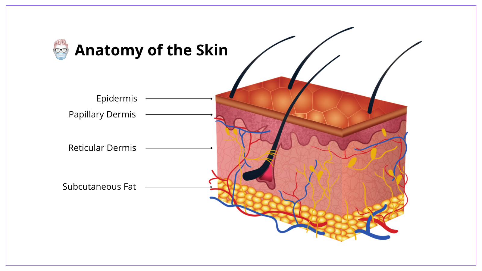
Anatomical Layers of the Skin
Epidermis
The skin's epidermis is stratified squamous epithelium and is derived from the ectoderm. The layers of the skin can be visualised in the image below.
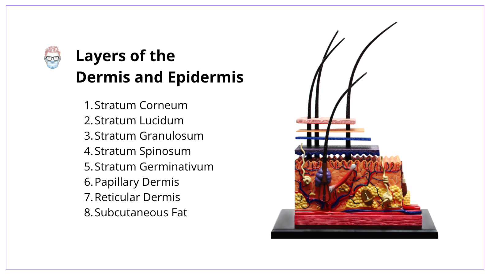
Within the epidermis, there are 5 layers:
- Stratum Germinativum: the deepest layer, contains melanocytes
- Stratum Spinosum: contains keratinocytes.
- Stratum Granulosum: contains mature keratinocytes.
- Stratum Lucidum: only present in glabrous skin.
- Stratum Corneum: contains non-viable keratinised cells.
The predominant cell type of the epidermis is keratinocytes. Other cells found in the epidermis include Melanocytes, Langerhans cells (APCs for the immune system) and Merkel Cells (neural crest mechanoreceptors).
Dermis
The dermis makes up most of the skin thickness and is derived from the mesoderm.
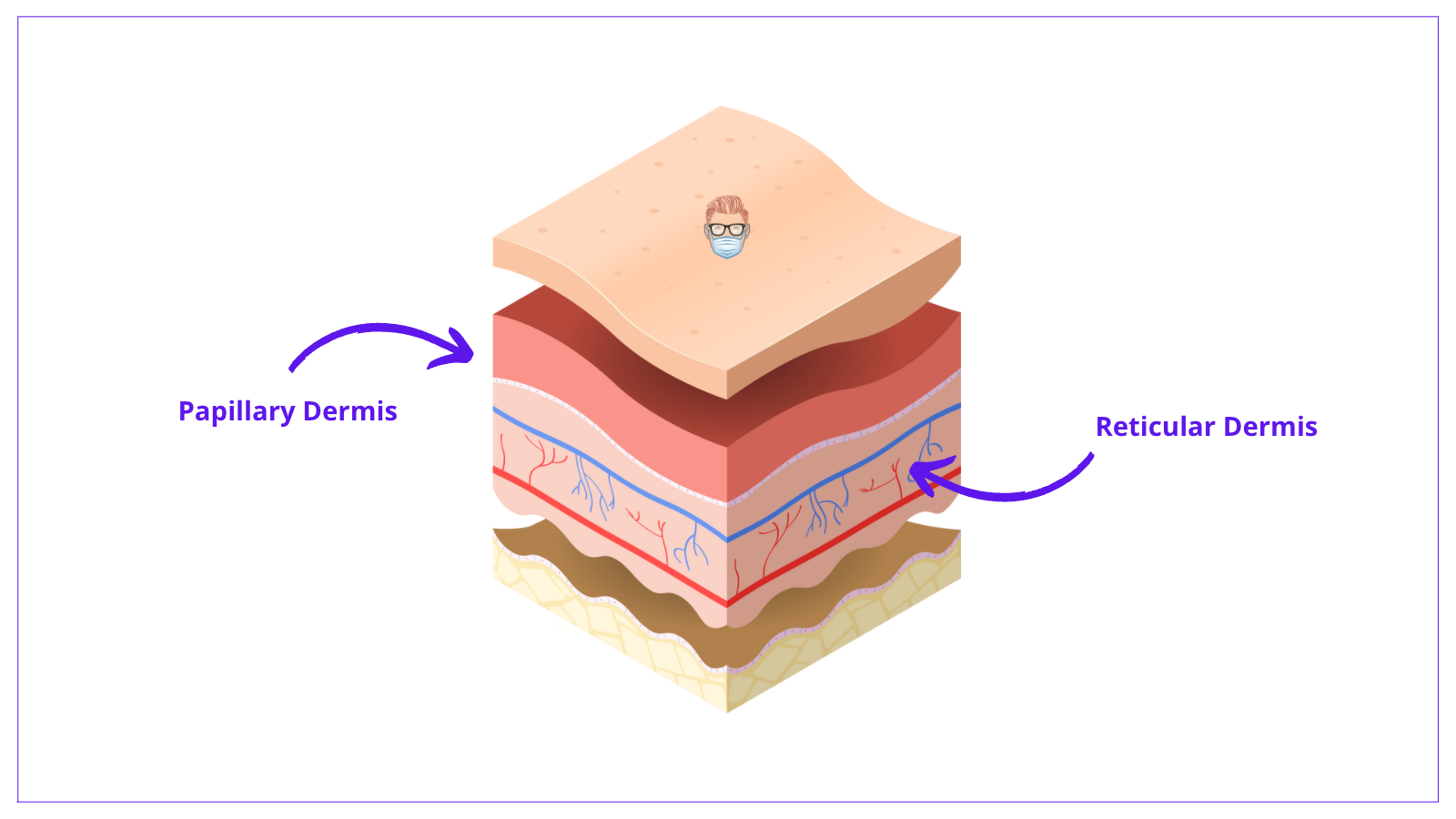
There are two layers in the dermis.
- Papillary dermis: superficial, fine collagen fibres, type III collagen.
- Reticular dermis: deeper, coarser collagen fibres, type I collagen.
The layers of the dermis can be seen in the image below.
The structures within the dermis are most commonly produced and secreted by fibroblasts. These include:
- Collagen: standard ratio of type 1: type 3 is 5:1 (some literature quotes 4:1). There are 5 different types of collagen. There is increased collagen III in ageing skin.
- Elastin: as the name suggests, it provides skin elasticity.
- "Ground Substance"/Glycosaminoglycans: hyaluronic acid, dermatan sulphate, chondroitin sulphate.
- Other cells include fibroblasts and mast cells.
Skin Appendages
Hair Follicles
- Arises from the epidermis and dermis of the skin.
- Drainage from sebaceous glands via the contraction of arrector pili muscles.
- It contains an inner root (from epidermis) and an outer root (from dermis).
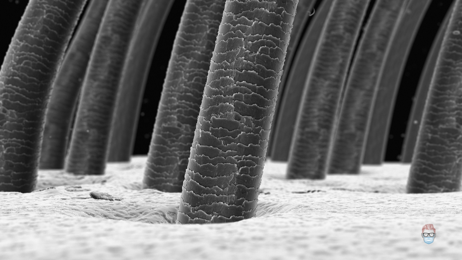
Hair
- Hair is composed of medulla, cortex and outer cuticle.
- 2 types of hair: fine vellus and coarse terminal hairs.
- They undergo a growth (anagen - most common), regressing (catagen - least common) and resting (telogen) phase.
Glands
There are three main glands in the skin: eccrine, apocrine and sebaceous
- Eccrine: odourless sweat glands in most locations in the body
- Apocrine: odour sweat glands, mainly found in the axilla and groin.
- Sebaceous: sebum-producing holocrine glands that drain in pilosebaceous units in hair-bearing skin and directly onto the skin in labia minor and penis.
Here is a video illustrating the role of these glands.
Other appendages include Meissner's corpuscle (light touch), Pacinian corpuscle (vibration, deep pressure), Bulb of Krause (cold temperature), Ruffini ending (hot temperature).
Ageing Skin Anatomy

Blood Supply to the Skin
It is relatively straightforward to understand the anatomical blood supply to the skin. There are 3 aspects to it: deep vessels, perforating vessels and plexuses. This is explained in the image below.
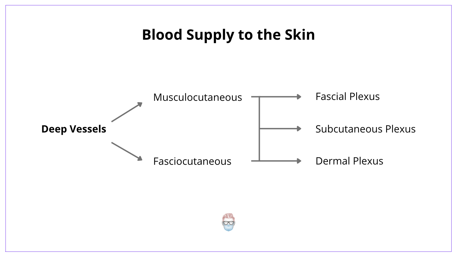
- Deep Vessels: arteries such as the aorta or common carotids etc
- Fasciocutaneous vessels: travel across the fascia directly to the skin
- Musculocutaneous vessels: travel in the muscle and indirectly provided perforators to the skin.
- Plexuses: there are a series of fascial, subcutaneous and dermal plexuses.
Active Recall Test
1. What is the most superficial layer of the skin?
The stratum corneum in the epidermis is the most superficial layer.
2. What is the deepest layer of the epidermis?
The stratum germinativum (also called basale). It is the origin of various skin cancers and contains melanocytes.
3. Name the most common collagen in the skin?
Type I collagen is the most common. It has a ratio of type 1:type III = 5:1. Type III collagen is increased in ageing and keloid scarring.
4. What is the embryological origin of the skin?
The epidermis arises from the ectoderm. The dermis arises from the mesoderm.
5. Where do hair follicles arise from?
They arise from the epidermis and dermis of the skin. They receive drainage from the sebaceous glands via the contraction of the erector pili muscles.
