In this Article on Bilobed Flaps
- 5 Key Points
- Definition of Bilobed Flap
- History of Bilobed Flaps
- Indications
- How to Design a Bilobed Flap
- Benefits vs Disadvantages
5 Key Points
- What is a bilobed flap? A Bilobed flap is classified as a random-pattern double transposition flap.
- Who created the bilobed flap? Originally described by Esser in 1918 and now the most commonly used is the 1989 Zitelli Modification
- How do you design a bilobed flap? By using the 3-2-1 rule to measure the geometry of the defect.
- When do you use it? The most common indication is nasal tip defects. There are several variations published and can also be used elsewhere.
- What are the specific complications? Pincushioning and alar deformation.
Definition of Bilobed Flap
The bilobed flap is classified as a random-pattern double transposition flap. A z-plasty is also a double-transposition flap.
The key principles to this repair are5,6,7:
- First lobe fills the primary defect, second lobe fills the secondary defect.
- Double transposition will distribute tension to wider area of skin.
- No specific blood supply (random pattern).
History of Bilobed Flaps
Since it's original description in 1918, the bilobed flap has undergone step-wise improvements in design to increase arc of rotation and indications. The Zitelli-modification is the most commonly used currently.
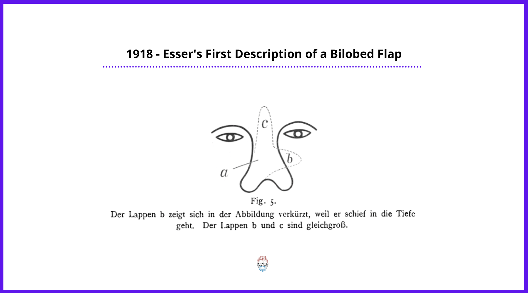
The bilobed flap was described by Esser in 191815.
- He used this flap for nasal tip reconstruction.
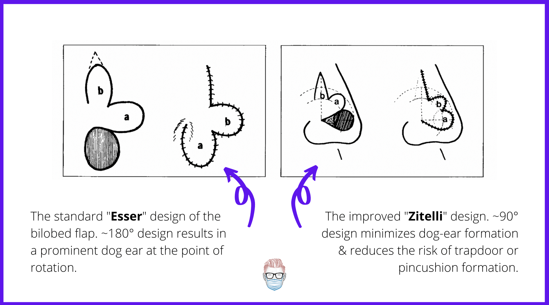
- Achieved 180° rotation with large dog-ear (see image above)
In 1953, Zimany modified the bilobed flap16.
- Demonstrated that the 2nd & 3rd lobes can be smaller than the defect.
- Showed flap could be utilized for many anatomical areas.
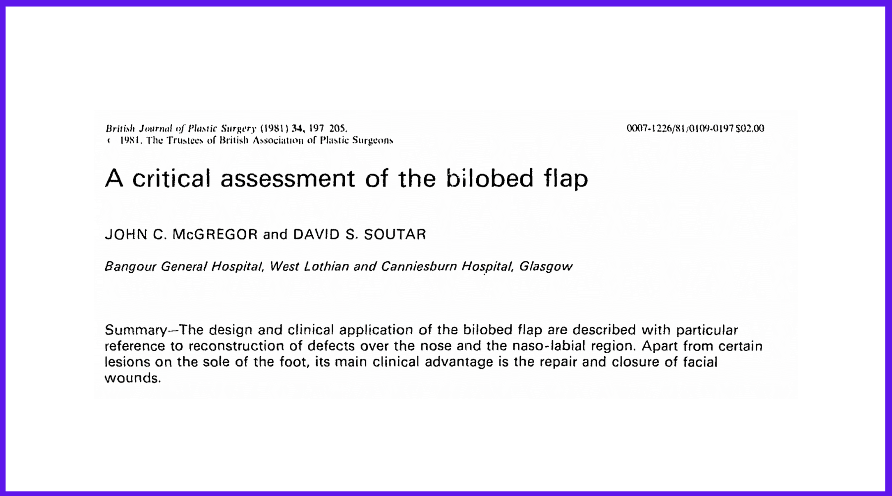
In 1980s, McGregor and Soutar13 further evaluated that by reducing the pivotal angle, the cutaneous deformity can be reduced. They also suggest the primary role should be in the face, rather than trunk and extremities.
Zitelli described the geometric bilobed flap which is mostly used nowadays which has limited arc of rotation up to 90 to 110 degrees14. The following illustration is adapted from Zitelli's original publication in 1989.
Indications for Bilobed Flap Repair
The are various indications for the bilobed flap. It is both patient, defect-size and surgeon-dependent.
The following are some general guidelines in relation to nose reconstruction8.
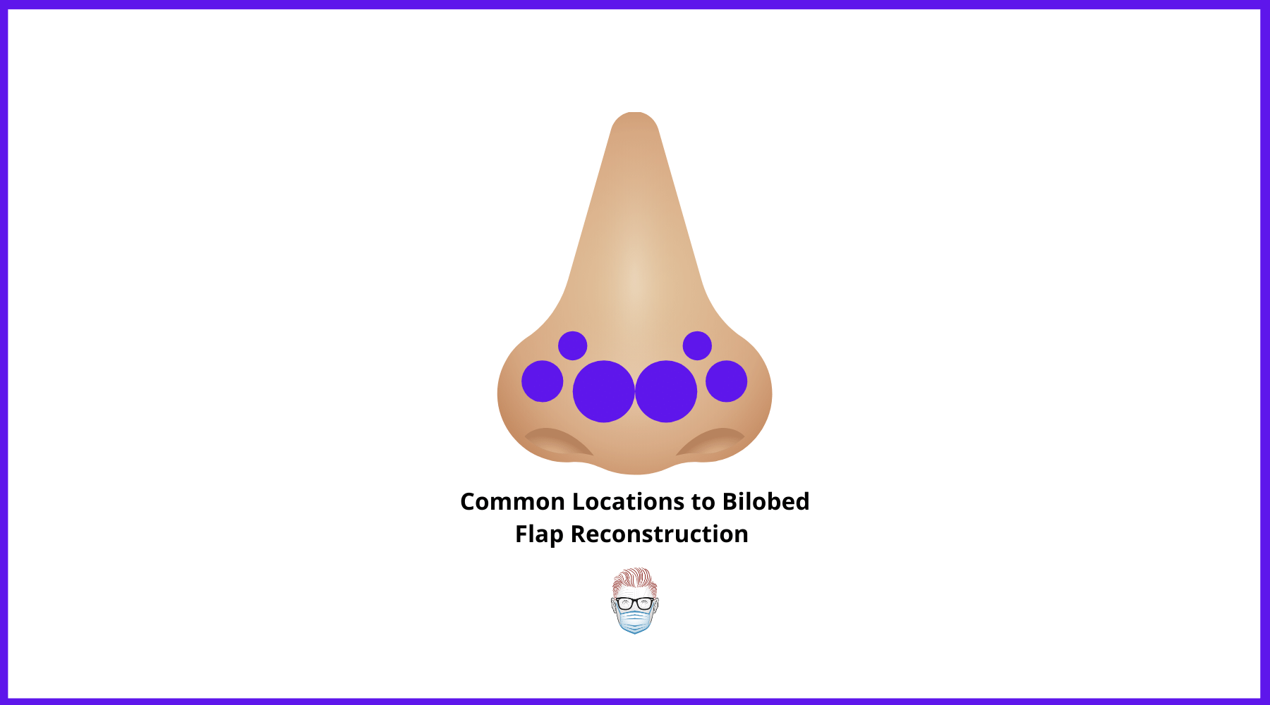
- Small to medium-sized cutaneous defects (0.5 to 1.5 cm)
- In the region of the inferior nose including nasal tip and ala.
- Defects should be more than 1 cm away from the nostril margin.
Interestingly, it also has a role in other areas of the body. For example, reconstruction post-resection of synovial cysts of the DIPJ. This can be read below.

How to Design a Bilobed Flap
The geometry of the bilobed flap can be difficult to design. This is because of the measurements, the contour changes of the nose, and also the fear of it not coming together! P'Fella has created this 3-2-1 mnemonic for bilobed flaps below.
The 3-2-1 Mnemonic
To simplify the geometry of the bilobed flap, just remember 3-2-1.
- 3 measurements: r, 2r, 3r
- 2 angles at 45-degrees
- 1 pivot point
By knowing these 3 steps, you have the foundation of how to draw a bilobed flap. This 3-2-1 rule is similar to Z-plasty design also. This is illustrated in the picture below.
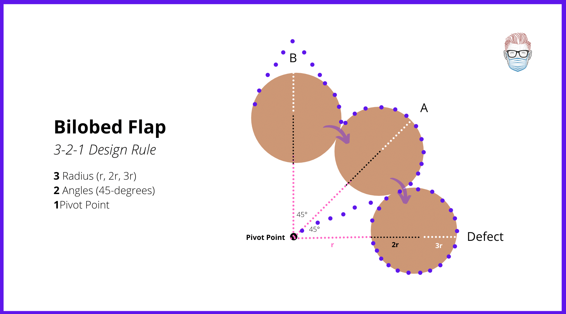
Steps to Designing Bilobed Flap
The following is a list of key landmarks required in a successful bilobed flap. This helps understand the geometry of the illustration above. Surgeons can raise this flap in a subcutaneous, submuscular, or periosteal plan. Variations do exist.
1. Defect Size
- This is the area that needs to be filled by the bilobed flap.
- The radius/diameter of the defect is the key to all future measurements.
2. Pivot Point
- A distance from the defect is equal to the radius' defect.
- The farther the pivot point is away from the defect, the larger the flap.
3. Lobule A
- At a 45-degree angle, the concentric circle is drawn at the same diameter and distance as the defect.
4. Lobule B
- At a 90-degree angle from the pivot point.
- This skin is often more mobile, so can be slightly smaller or in a more triangular fashion.
5. Dog-Ear/SCD
- A dog-ear excision is marked from the defect to the pivot point
- This creates space for the flap to rotate and advance.
Some technical points to consider during the operation are:
- Defect B (donor site) is closed first.
- Thin the lobes to match the contour of that area of the nose.
- Pincushioning can be avoided if it is carefully repaired in layers.
- Consider the topographic nasal subunit principle of Burget and Menick2,3,4
Discussion
Benefits
- Allows movement of more skin over a larger distance than would be possible with a single transposition flap (for example, a rhomboid flap)
- Fills defects with nearby skin that is matched for color and texture3.
- Reliable and versatile reconstruction for noses1
Disadvantages
- Dog-Ear Formations. The conventional bilobed flap gives a large dog ear, which when removed, can narrow the base of the flap and reduce its blood supply6
- Pincushioning and distortion of alar margins are recognized complications, in addition to the standard surgical complications9.
Modifications
- Superiorly-based bilobed flap moves the terminal donor site of the nose and decreases the risk for lower nasal distortion. The superiorly based bilobed flap preserves the advantages of the traditional design and expands the utility of this flap8.
- Dual rhombic flaps in order to decrease the incidence of flap pincushioning from other nasal flap designs, including the standard bilobed flap10
- Bilobed islanded flap can increase the rotation up to 180° and resolve the issue of dog ears without hampering the blood supply of the flap. In this modification, flap lobes are islanded on the transverse part of the nasalis muscle supplied by the external nasal artery11
References:
1. Salgarelli AC, Bellini P, Multinu A, Magnoni C, Francomano M, Fantini F, Consolo U, Seidenari S. Reconstruction of nasal skin cancer defects with local flaps. Journal of Skin Cancer. 2011 Jan; 181093:01-08.
2. Maher IA. Extrapolating Straight Lines to Curves: Can the Dynamics of Z-Plasties Be Applied to Bilobed and Trilobed Flaps? Dermatol Surg. 2020 Feb;46(2):277-280.
3. Kim YH, Yoon HW, Chung S, Chung YK. Reconstruction of cutaneous defects of the nasal tip and alar by two different methods. Arch Craniofac Surg. 2018 Dec;19(4):260-263.
4. Filitis DC, Fisher J, Samie FH. Reconstruction of a surgical defect in the popliteal fossa: A case report. Int J Surg Case Rep. 2018;53:228-230.
5. Ramanujam CL, Zgonis T. Use of Local Flaps for Soft-Tissue Closure in Diabetic Foot Wounds: A Systematic Review. Foot Ankle Spec. 2019 Jun;12(3):286-293.
6. Knackstedt T, Lee K, Jellinek NJ. The Differential Use of Bilobed and Trilobed Transposition Flaps in Cutaneous Nasal Reconstructive Surgery. Plast Reconstr Surg. 2018 Aug;142(2):511-519.
7. Grieco MP, Bertozzi N, Grignaffini E, Raposio E. Nose defects reconstruction with the Zitelli bilobed flap. G Ital Dermatol Venereol. 2018 Apr;153(2):278-282.
8. Kelly-Sell M, Hollmig ST, Cook J. The superiorly based bilobed flap for nasal reconstruction. J Am Acad Dermatol. 2018 Feb;78(2):370-376.
9. Menick FJ. Aesthetic nasal reconstruction. Neligan PC. Plastic Surgery. 4th edi. Canada: Elsevier Inc;2018;03:135-137.
10. Dinehart SM. The rhombic bilobed flap for nasal reconstruction. Dermatol Surg 2001;27:501-504.
11. Goldman R, Speranzini MB, Goldman B. The bilobed island flap in nasal ala reconstruction. British Journal of Plastic Surgery. 1998;51:493-498.
12. Zoumalan RA, Hasan C, Levine VJ, Shah AR. Analysis of vector alignment with the Zitelli bilobed flap for nasal defect repair: a comparison of flap dynamics in human cadavers. Archives of facial plastic surgery. 2008;10(3):181-185.
13. JOHN C. McGREGOR and DAVID S. SOUTAR, 1981. A critical assessment of the bilobed flap. British Journal of Plastic Surgery. 0007-1226/81/0109-(1197$02.00
14. Zitelli, J. A. (1989). The Bilobed Flap for Nasal Reconstruction. Archives of Dermatology, 125(7), 957.doi:10.1001/archderm.1989.01670190091012
15. Esser JF. Gestielte loakle Nasenplastik mit zweizipfligen Lappen, Deckung des sekundaren Defektes vom ersten Zipfel durch den Zweiten. Deutsche Zeitschrift für Chirurgie. 1918;143:335-9.
16. Zimany A. The Bi-Lobed Flap. Plast Recon Surg. 1952 Jun;11(6):424-34.
Authors: Dr. Darshil Rajgor, SCL Municipal General Hospital, Saraspur, Ahmedabad, Gujarat, India.
Mentors: Prof. Nilesh B. Ghelani, Associate Prof. Sankit D. Shah
Verified by thePlasticsFella
Post Publication Editors:
- Dr Juan Varela, A Coruña, Spain: provided information regarding the role of bilobed flaps in finger reconstructions.


