


Brow assessment is necessary because heavy upper eyelids, tired appearance, and superior visual field symptoms may reflect brow ptosis, dermatochalasis, true blepharoptosis, or a combination.
Periorbital Unit Assessment
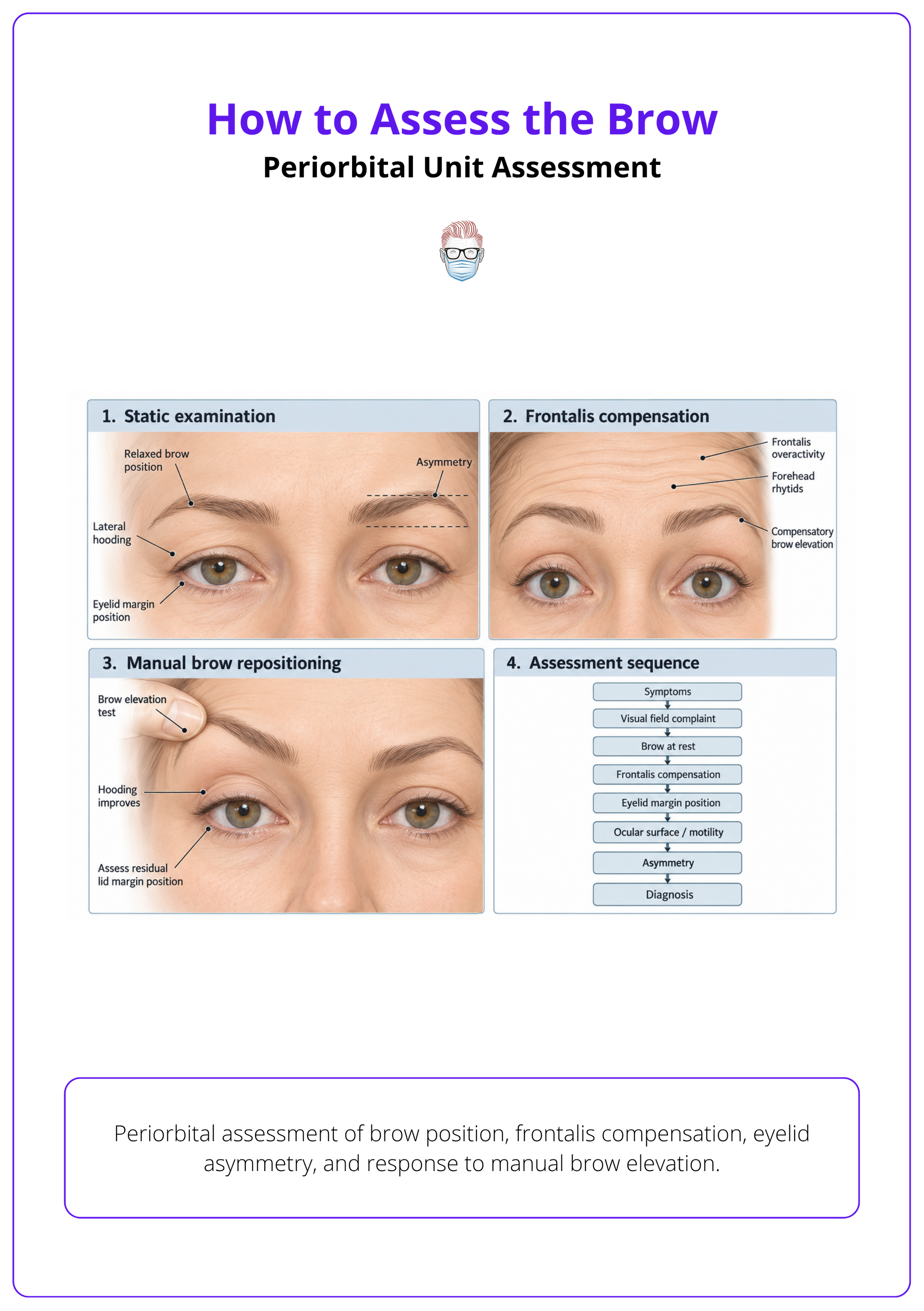
Assess the brow in sequence: symptoms, superior visual field defects, apparent dermatochalasis, relaxed brow position, frontalis compensation, eyelid margin position, ocular function, and facial asymmetry.
Applied Brow Anatomy
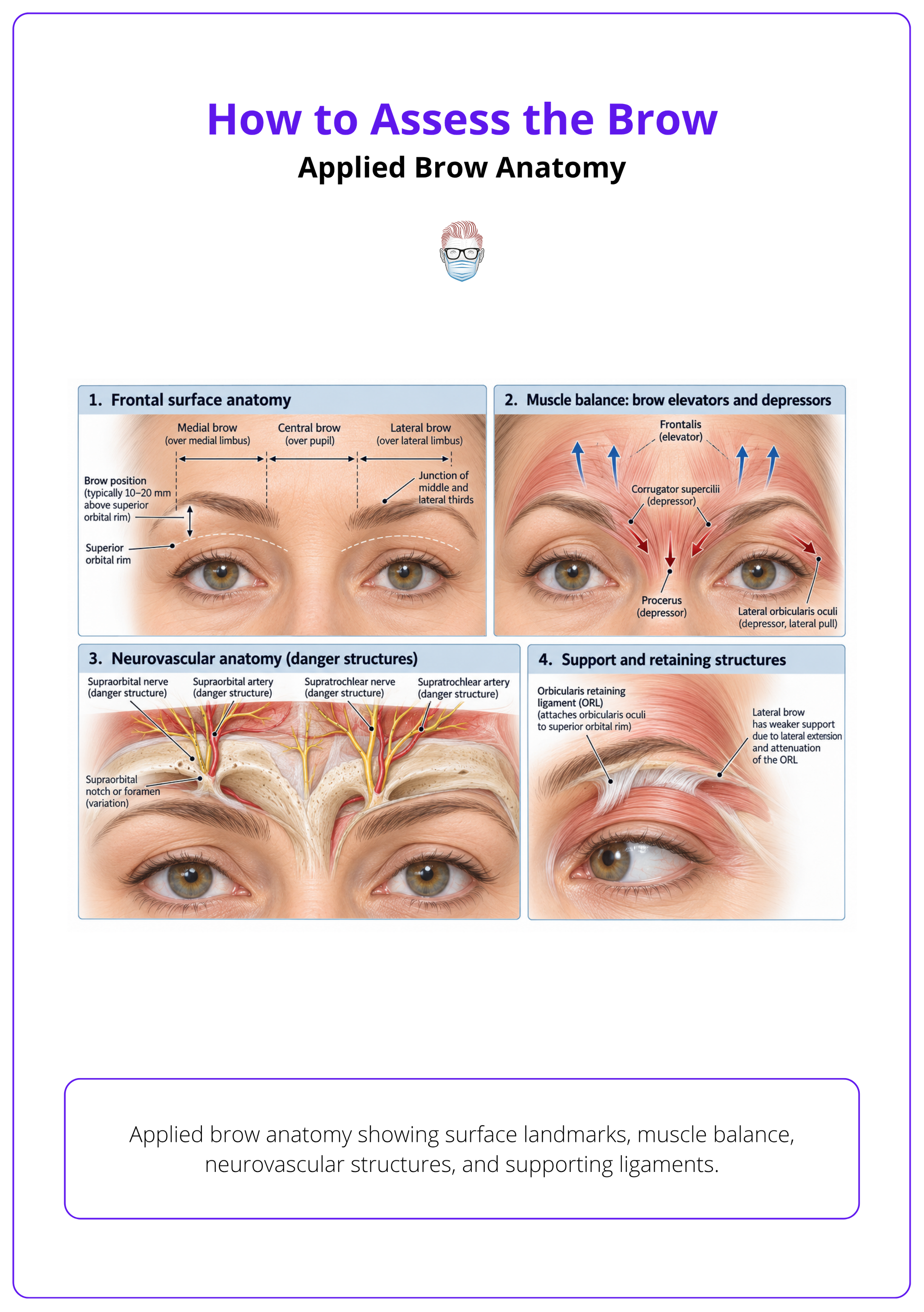
The anatomy that changes brow assessment is the brow relationship to the orbital rim, sex- and age-related brow morphology, frontalis and depressor balance, and supraorbital neurovascular danger structures.
Objective Measurements and Classification
Use resting brow position, brow height landmarks, asymmetry, margin reflex distance-1, levator function, dermatochalasis grading, and standardised photographs to separate brow ptosis from eyelid pathology.
Differential Diagnosis
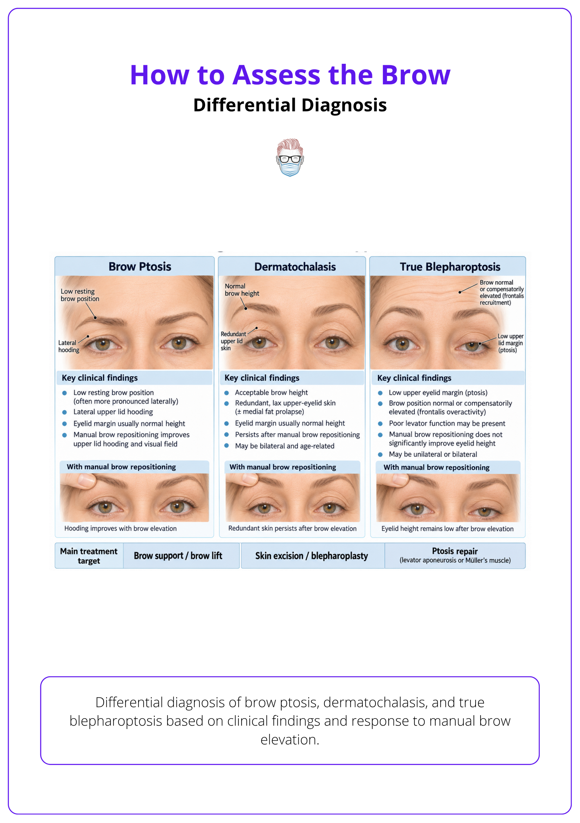
Brow ptosis improves when the brow is repositioned, dermatochalasis persists as redundant eyelid skin with acceptable brow height, and true blepharoptosis persists as a low eyelid margin after brow correction.
Risk Assessment and Investigations
Safety checks should identify ocular surface risk, exposure keratopathy risk, facial palsy, thyroid eye disease, prior refractive or periocular procedures, botulinum toxin-related brow or lid changes, and functional visual field defects.
Treatment Planning
Translate assessment into treatment by treating the dominant problem: observe mild asymptomatic descent, use non-operative options for selected mild dynamic or laxity-driven cases, lift the brow for true brow ptosis, repair the eyelid for true blepharoptosis, and combine procedures when pathology is mixed.
Clinical Documentation and Visual Aids
Documentation should make brow assessment reproducible by recording standardised photographs, landmark measurements, visual field evidence, frontalis compensation, and baseline brow–eyelid relationships.
Clinical Framing
Brow assessment is necessary because heavy upper eyelids, tired appearance, and superior visual field symptoms may reflect brow ptosis, dermatochalasis, true blepharoptosis, or a combination.
Patients rarely present saying “my brow is ptotic.” They usually describe heavy lids, excess upper-eyelid skin, visual field defects, or a tired look.
Diagnostic Purpose
Start by deciding whether the apparent upper-lid problem is brow-related, eyelid-skin related, eyelid-margin related, or mixed.
- Apparent dermatochalasis: upper-eyelid laxity should prompt brow assessment, not just eyelid-skin planning 1.
- Visual field defects: assessment must consider functional visual interference, not only cosmetic appearance 2.
- Tired appearance: brow position should be assessed early because brow descent affects upper-face aesthetics 1.
- Symptom burden: document perceived functional impairment when brow and dermatochalasis coexist 3.
Brow-Lid Unit
Assess the eyebrow and upper eyelid as one functional unit before offering blepharoplasty, brow lift, or ptosis repair.
- Periorbital unit: evaluate the brow within the eyelid-eyebrow complex, not as an isolated eyebrow height problem 4.
- Blepharoplasty trap: counsel that upper-eyelid surgery can lower brow position, so brow assessment must precede blepharoplasty planning 5.
- Unaddressed descent: clinically relevant brow ptosis may compromise the eyelid surgical result if missed 6.
- Baseline asymmetry: identify pre-existing brow asymmetry before attributing it to treatment or complication 2.
Periorbital Unit Assessment
Assess the brow in sequence: symptoms, superior visual field defects, apparent dermatochalasis, relaxed brow position, frontalis compensation, eyelid margin position, ocular function, and facial asymmetry.
The brow is assessed in the context of periorbital ageing, eyelid function, facial animation, and patient expectation. A relaxed, neutral examination prevents frontalis overactivity from hiding brow ptosis.
History
Let the presenting symptom guide the diagnostic pathway without assuming the problem is isolated eyelid skin.
- Common symptoms: heaviness, upper-eyelid excess, superior visual field loss, tired appearance, headache, or frontalis fatigue.
- Aesthetic concerns: record brow shape, lateral hooding, eyelid fold change, wrinkles, and age-related expectations 1.
- Botulinum toxin type A: document prior or planned BoNT-A pattern because the injection technique changes brow elevation 7.
- Forehead treatment: evaluate forehead rhytids and brow height together when interpreting BoNT-A effects 8.
Static Examination
Examine in primary gaze with the brows relaxed so brow descent and asymmetry are not masked.
- Neutral position: assess brow and eyelids in relaxed primary gaze to avoid masking ptosis or asymmetry 2.
- Apparent dermatochalasis: upper-eyelid laxity should prompt assessment of brow position, not just eyelid skin 1.
- Facial asymmetry: include asymmetry and patient expectations in the baseline brow assessment 4.
- Baseline comparator: eyelid procedures may be followed by measurable brow descent, so preoperative brow position matters 5.
Dynamic Examination
Separate active frontalis elevation from true resting brow position.
- Frontalis compensation: look for forehead rhytids, raised brows, and brow fatigue from chronic recruitment.
- Relaxation test: manually block or cue frontalis relaxation before judging true brow height.
- Depressor activity: observe glabellar and orbicularis recruitment, especially in lateral hooding.
- Ocular motility: include extraocular muscle assessment when lid elevation is abnormal or toxin spread is suspected 2.
Ocular Examination
Identify ocular findings that change whether brow, eyelid, combined, or referral-based management is safe.
- Visual baseline: check visual acuity, consider visual fields, and assess ocular motility before rejuvenation planning 4.
- Dry eye symptoms: screen before brow or periorbital surgery because pre-existing dry eye may increase postoperative risk 4.
- Ocular prominence: Hertel values over 18 mm may indicate higher risk of lid malposition, scleral show, or dry eye 4.
- Facial paralysis: document eyelid closure gap and MRD2 when periorbital function and symmetry are affected 9.
Applied Brow Anatomy
The anatomy that changes brow assessment is the brow relationship to the orbital rim, sex- and age-related brow morphology, frontalis and depressor balance, and supraorbital neurovascular danger structures.
Anatomy is useful only if it changes diagnosis, planning, or risk. For brow assessment, the clinically relevant question is whether the brow is low, weakly supported, asymmetrical, or unsafe to treat casually.
Surface Anatomy
Judge brow position against the orbital rim, contour, and patient-specific norms rather than a single ideal brow.
- Female brow: young female brows are commonly described around 5 mm above the orbital margin 1.
- Male brow: male brows are often assessed around the orbital rim rather than high above it 1.
- Patient variation: adjust targets for gender, age, and ethnicity rather than applying one brow ideal 4.
- Contour reference: the junction of the middle and lateral thirds helps judge brow shape and lift goals 4.
Muscle Anatomy
Brow position reflects the balance between frontalis elevation and orbicularis-glabellar depressor activity.
- Frontalis: chronic recruitment can hide brow ptosis and create compensatory forehead lines.
- Orbicularis: lateral orbicularis descent contributes to lateral hooding and crow’s-feet patterning.
- Depressor mapping: separate medial and lateral depressor function because each produces different brow-shaping effects 7.
- Dynamic assessment: evaluate frontalis elevation and depressor activity during planning 4.
Neurovascular Anatomy
Identify sensory and vascular structures before injection, energy treatment, or operative planning.
- Supraorbital exit: notch versus foramen variation has been reported at about 74% and 26% respectively 2.
- Superior pedicles: supraorbital and supratrochlear pathways can transmit unintended toxin spread 2.
- Energy mapping: upper-face ultrasound planning should avoid or minimise exposure over supraorbital and zygomatic nerve pathways 1.
- Retaining ligament: account for the orbicularis retaining ligament during brow-lift planning to reduce ocular risk 4.
Objective Measurements and Classification
Use resting brow position, brow height landmarks, asymmetry, margin reflex distance-1, levator function, dermatochalasis grading, and standardised photographs to separate brow ptosis from eyelid pathology.
Objective measurements do not replace clinical judgement. They make the diagnosis reproducible, support functional documentation, and prevent confusing brow descent with eyelid-margin ptosis.
Brow Measurements
Measure the brow at rest using reproducible landmarks and without frontalis or glabellar contraction.
- True rest: measure brow ptosis with the brow relaxed, eyes closed, and no frontalis or glabellar contraction 4.
- Baseline line: a horizontal line connecting the medial canthi can provide a measurement baseline 1.
- Vertical landmarks: brow height can be measured through canthi, iris edges, and pupil centre 1.
- Brow edge: record vertical distance from canthal baseline to the upper eyebrow edge at standardised points 1.
| Measurement | How to Record It | Diagnostic Use |
|---|---|---|
| Brow height | Vertical brow distance at standardised landmarks | Tracks descent and asymmetry 1 |
| Orbital rim relationship | Brow below superior orbital rim or <10 mm above lid margin | Supports brow ptosis 4 |
| Male mid-pupil distance | Mid-pupil to top-of-brow distance | <2.5 cm suggests brow ptosis in men 4 |
| Margin reflex distance-1 (MRD-1) | Corneal light reflex to upper lid margin | Normal described as 4.0–4.5 mm 2 |
| Levator function | Excursion with frontalis held still | ≥14 mm stated as normal 2 |
Eyelid Measurements
Measure the eyelid because a low lid margin position changes the diagnosis and treatment.
- MRD-1 definition: margin reflex distance-1 is the distance from the corneal light reflex to the upper eyelid margin.
- Levator function: hold the frontalis still when measuring excursion to avoid overestimating lid power 2.
- Palpebral fissure: measure at the mid-pupillary line when grading eyelid opening 2.
- Eyelid fold height: use eyelid fold height or MRD-1 to distinguish eyelid ptosis from brow descent 4.
Severity Classification
Classify severity so the diagnosis, documentation, communication, and procedure choice align.
- Structured grading: use a brow ptosis classification to guide diagnosis, documentation, and procedure selection 10.
- Brow ptosis: classify when the brow falls below the superior orbital rim or sits <10 mm above the eyelid margin 4.
- Fissure grading: palpebral fissure deficit may be graded mild 1–2 mm, moderate 3–4 mm, or severe >4 mm 2.
- Asymmetry: record side-to-side difference separately from absolute brow height.
Photographic Measurement
Use photographs to support serial comparison, not to replace examination.
- Standardised images: measure vertical brow displacement in millimetres on standardised photographs when objective change matters 11.
- Neutral expression: capture relaxed brow, gentle eye closure, and primary gaze.
- Dynamic views: include forced brow elevation and frontalis relaxation when compensation is clinically relevant.
- Decision value: photographs should show whether manual brow repositioning corrects hooding or unmasks eyelid ptosis.
Differential Diagnosis
Brow ptosis improves when the brow is repositioned, dermatochalasis persists as redundant eyelid skin with acceptable brow height, and true blepharoptosis persists as a low eyelid margin after brow correction.
This is the central diagnostic task. Do not book “upper blepharoplasty” until you know whether the brow, eyelid skin, lid margin, or another disease process is driving the complaint.
Diagnostic Comparison
Compare brow position, eyelid skin, lid margin, and frontalis recruitment in the same examination.
| Finding | Brow Ptosis | Dermatochalasis | True Blepharoptosis |
|---|---|---|---|
| Brow position | Low at rest, often lateral | Acceptable | May be normal or compensatory high |
| Upper-lid fold | Low sulcus may suggest brow descent 4 | Redundant skin fold | Lid margin remains low |
| Manual brow lift | Hooding improves | Skin excess persists | Ptosis persists |
| Frontalis use | Often recruited | Variable | Often recruited |
| Main treatment target | Brow support | Skin excision | Ptosis repair |
Apparent Dermatochalasis
Treat lateral hooding as a brow diagnosis until proven otherwise.
- Lateral hooding: assess for lateral brow ptosis before treating it as isolated upper-eyelid skin excess 4.
- Low sulcus: a low upper lid sulcus can indicate that apparent skin excess originates from brow descent 4.
- Lacrimal fullness: consider prolapsed lacrimal gland when lateral upper-eyelid fullness is present 4.
- Skin-only excess: true dermatochalasis has an acceptable brow position and lid margin, with redundant eyelid skin.
Blepharoptosis
A low eyelid margin is eyelid ptosis until the levator and ptosis category prove otherwise.
- Ptosis categories: classify drooping upper lid as neurogenic, myogenic, aponeurotic, mechanical, or traumatic 2.
- Acquired causes: distinguish involutional and congenital ptosis from neurogenic, myogenic, and posttraumatic causes 12.
- Congenital ptosis: eyelid droop from birth is not primary eyebrow descent 13.
- Mass lesion: unilateral acquired ptosis with a hard upper-lid mass warrants systemic or malignant evaluation 14.
Secondary Causes
Name the cause of brow or lid descent before planning aesthetic correction.
- Neurologic causes: include myasthenia gravis, facial paralysis, trauma, and congenital or hereditary causes 1.
- Horner’s syndrome: consider acquired causes such as Horner’s syndrome and myasthenia gravis in apparent droop 2.
- Botulinum toxin changes: recent forehead BoNT-A pattern can contribute to brow-position change 8.
- Postoperative descent: prior ptosis correction may contribute to brow descent and should not be mistaken for primary brow ptosis 5.
Risk Assessment and Investigations
Safety checks should identify ocular surface risk, exposure keratopathy risk, facial palsy, thyroid eye disease, prior refractive or periocular procedures, botulinum toxin-related brow or lid changes, and functional visual field defects.
Risk assessment determines whether to proceed, defer, modify the plan, or refer. The aim is not to order every test, but to find the risk that changes management.
Ocular Surface
Protect the cornea before planning brow, eyelid, or combined intervention.
- Ocular risk: dry eye, glaucoma, contact lens use, trauma, allergies, epiphora, and previous ocular surgery.
- Exposure signs: lagophthalmos, weak Bell’s phenomenon, scleral show, epithelial compromise, or exposure keratopathy.
- Surface testing: use Schirmer testing, tear break-up time, staining, and tear meniscus height when ocular surface risk changes planning 4.
- Refractive surgery: delay blepharoplasty-related planning until 6 months after refractive surgery to reduce ocular surface injury risk 4.
Neuromuscular History
Identify neuromuscular, thyroid, and facial nerve pathology before attributing asymmetry to ageing.
- Facial weakness: facial palsy, Bell’s palsy, incomplete closure, synkinesis, or previous induced eyelid ptosis 2.
- Medication risk: neuromuscular disease and drugs such as aminoglycosides, warfarin, and Alzheimer’s medications may affect BoNT-A planning 2.
- Thyroid dysfunction: thyroid disease affects suitability and planning for periorbital surgery 4.
- Thyroid eye disease: defer rehabilitative brow or eyelid surgery until quiescence is documented for at least 6-9 months 4.
Botulinum Toxin Changes
Distinguish botulinum toxin type A (BoNT-A)-related brow ptosis from eyelid ptosis before offering surgical correction.
- Brow versus lid: document whether post-glabellar BoNT-A ptosis is eyebrow descent or eyelid-margin ptosis 15.
- Event counselling: repeated glabellar BoNT-A series reported 55 ptosis events in 45 of 1200 patients, with most episodes lasting less than 3 weeks 15.
- Timing: correlate onset with injection date, pattern, dose, and any change in frontalis recruitment.
- Escalation: atypical diplopia, anisocoria, new neurologic signs, or persistent unilateral ptosis needs ophthalmic or neurologic review.
Investigations
Photography
Photograph the brow before examination, manipulation, toxin, or surgery changes its resting position.
- Visual field defects: document the cause of upper-field impairment before deciding on brow, eyelid, or combined surgery 16.
- Functional planning: include anaesthesia planning, need for adjunctive brow procedure, and follow-up strategy in functional upper-field cases 16.
- Microfocused ultrasound: screen MFU candidates for excessive laxity and BMI >30 because these were treated as relative contraindications 11.
- Energy-based treatment: screen for pregnancy, systemic disease, active skin disease, infection, sensory or motor disorders, and follow-up reliability 1.
Treatment Planning
Translate assessment into treatment by treating the dominant problem: observe mild asymptomatic descent, use non-operative options for selected mild dynamic or laxity-driven cases, lift the brow for true brow ptosis, repair the eyelid for true blepharoptosis, and combine procedures when pathology is mixed.
Treatment planning follows diagnosis. Do not use blepharoplasty to solve a low brow, and do not use a brow lift to solve a low eyelid margin.
Decision Sequence
Choose the intervention that matches the anatomical source of heaviness or obstruction.
| Assessment Finding | Best-Fit Plan | Main Pitfall |
|---|---|---|
| Mild asymptomatic descent | Observation, photographs, review | Overtreatment |
| Dynamic depressor dominance | BoNT-A brow shaping | Overweakening frontalis |
| Mild–moderate laxity and brow ptosis | Consider microfocused ultrasound | Millimetric lift only 1 |
| Severe brow ptosis | Surgical brow lift | Expecting non-operative correction 1 |
| Dermatochalasis plus brow descent | Combined brow lift and blepharoplasty | Treating eyelid skin alone 4 |
| True blepharoptosis | Ptosis repair | Missing levator pathology |
Non-Operative Treatment
Use non-operative treatment only when the deformity is mild, dynamic, reversible, or skin-quality driven.
- BoNT-A planning: upper forehead placement may reduce brow-ptosis risk but gives less forehead-line improvement than lower patterns 8.
- Injection height: middle-forehead BoNT-A placement lowered the eyebrow more than upper-forehead placement 8.
- Safety margins: glabellar BoNT-A planning should place the injection 1 cm above the brow to reduce eyelid ptosis risk 2.
- MFU counselling: microfocused ultrasound is an energy-based tightening option for selected mild to moderate brow ptosis, not a substitute for surgical elevation 1.
Non-Operative Outcomes
Counsel non-operative brow lift in millimetres rather than dramatic surgical repositioning.
- MFU response: in mild upper-eyelid laxity and brow ptosis, 35 of 40 subjects were judged to have clinically significant brow elevation at 180 days 1.
- MFU magnitude: mean eyebrow elevation was about 2 mm at 90 and 180 days in the completed cohort 1.
- Crow’s-feet injections: avoid injections medial to the mid-pupillary line and keep at least 1 cm from the orbital margin inferiorly 2.
- Botulinum toxin timing: wait for the clinical pattern to declare itself before labelling asymmetry as structural brow ptosis.
Operative Selection
Use surgery when brow descent is structural, severe, functionally significant, or combined with eyelid pathology.
- Avoid isolated eyelid surgery: significant brow ptosis should be addressed because blepharoplasty or ptosis repair may further lower the brow 4.
- Combined pathology: plan combined brow lift and blepharoplasty when both brow descent and upper-eyelid excess contribute 4.
- Approach choice: do not reject open brow-lift approaches solely because endoscopic surgery is assumed superior 17.
- Evidence limits: explain that randomized comparative outcome data for brow-lift approaches were not available in the cited review 17.
Clinical Documentation and Visual Aids
Documentation should make brow assessment reproducible by recording standardised photographs, landmark measurements, visual field evidence, frontalis compensation, and baseline brow–eyelid relationships.
Good documentation protects the patient and surgeon. It also lets the plan be reviewed when symptoms, asymmetry, or postoperative change are disputed.
Clinical Photography
Photograph the brow and eyelids before any intervention changes position, tone, or compensation.
- Baseline images: obtain pre-treatment photographs as a visual aid for brow and eyelid assessment 2.
- Standard views: include frontal and bilateral oblique or profile photographs when assessing treatment effect 1.
- Defined landmarks: document brow position with standard landmarks and photography to support reproducible planning 18.
- 3D imaging: vector imaging can document direction and magnitude of brow and forehead skin displacement over time 1.
Measurement Diagrams
Use diagrams and templates to show what was measured, why it mattered, and how it changed the plan.
- Template consistency: use a uniform measurement and examination template for preoperative planning and postoperative comparison 4.
- Baseline comparison: record pre-treatment brow and eyelid position so outcomes or complications can be compared objectively 2.
- Functional cases: document the cause of visual field impairment and the rationale for selected repair 16.
- Teaching images: include examples of frontalis compensation, manual brow repositioning, brow ptosis, dermatochalasis, and true eyelid ptosis.
References
[1] Chen et al.. Ultrasound rejuvenation for upper facial skin: A randomized blinded prospective study.. Journal of cosmetic dermatology. 2024. doi:10.1111/jocd.16482
[2] Nestor et al.. Botulinum toxin-induced blepharoptosis: Anatomy, etiology, prevention, and therapeutic options.. Journal of cosmetic dermatology. 2021. doi:10.1111/jocd.14361
[3] Lee et al.. Is Blepharoplasty Cost-effective? Utility Analysis of Dermatochalasis and Cost-effectiveness Analysis of Upper Eyelid Blepharoplasty.. Ophthalmic plastic and reconstructive surgery. 2024. doi:10.1097/IOP.0000000000002649
[4] Kashkouli et al.. Periorbital facial rejuvenation; applied anatomy and pre-operative assessment.. Journal of current ophthalmology. 2017. doi:10.1016/j.joco.2017.04.001
[5] Liu et al.. Brow Position Change and its Potential Risk Factors Following Upper Blepharoplasty: A Systematic Review and Meta-Analysis.. Aesthetic plastic surgery. 2023. doi:10.1007/s00266-023-03288-0
[6] Sugino et al.. Comparison of two techniques for surgical eyebrow suspension.. Journal francais d'ophtalmologie. 2022. doi:10.1016/j.jfo.2022.03.011
[7] El-Khoury et al.. The Impact of Botulinum Toxin on Brow Height and Morphology: A Randomized Controlled Trial.. Plastic and reconstructive surgery. 2018. doi:10.1097/PRS.0000000000003919
[8] Jabbour et al.. The Impact of Upper Face Botulinum Toxin Injections on Eyebrow Height and Forehead Lines: A Randomized Controlled Trial and an Algorithmic Approach to Forehead Injection.. Plastic and reconstructive surgery. 2018. doi:10.1097/PRS.0000000000004836
[9] Zhou et al.. Enhancing Lower Eyelid Suspension Outcomes Through Pre-surgical Facial Nerve Reanimation: A Comparative Study.. Aesthetic plastic surgery. 2024. doi:10.1007/s00266-024-04241-5
[10] Pinheiro et al.. Clinical classification of brow ptosis. Revista Brasileira de Cirurgia Plástica. 2016. doi:10.5935/2177-1235.2016RBCP0058
[11] Contini et al.. A Systematic Review of the Efficacy of Microfocused Ultrasound for Facial Skin Tightening.. International journal of environmental research and public health. 2023. doi:10.3390/ijerph20021522
[12] De et al.. Blepharoptosis.. Minerva chirurgica. 2013. PMID:24172762
[13] Rosenberg et al.. Types of materials for frontalis sling surgery for congenital ptosis.. The Cochrane database of systematic reviews. 2019. doi:10.1002/14651858.CD012725.pub2
[14] Kinori et al.. Ptosis Shmosis.. Survey of ophthalmology. 2017. doi:10.1016/j.survophthal.2016.02.004
[15] Moy et al.. Long-term safety and efficacy of a new botulinum toxin type A in treating glabellar lines.. Archives of facial plastic surgery. 2009. doi:10.1001/archfacial.2009.5
[16] Kim et al.. American Society of Plastic Surgeons Evidence-Based Clinical Practice Guideline: Eyelid Surgery for Upper Visual Field Improvement.. Plastic and reconstructive surgery. 2022. doi:10.1097/PRS.0000000000009329
[17] Graham et al.. Brow lift in facial rejuvenation: a systematic literature review of open versus endoscopic techniques.. Plastic and reconstructive surgery. 2011. doi:10.1097/PRS.0b013e3182268d41
[18] Freitag et al.. Anthropometric analysis and aesthetic guidelines for eyebrow position: a review. Ophthalmic Plastic and Reconstructive Surgery. 2015. doi:10.1097/IOP.0000000000000450
