


Summary Card
Applied Eyelid Anatomy
Assess the eyelid by lamellae, margin, canthal support, levator function, orbicularis closure, lacrimal drainage, and corneal protection mechanisms because each changes diagnosis, risk, and treatment planning.
Lamellar Eyelid Examination
Examine the eyelid by anterior lamella, middle lamella, posterior lamella, and orbital support so that visible signs map to the layer causing the deformity.
History and Symptoms
Focus the history on onset, fluctuation, pain, vision, diplopia, exposure symptoms, tearing, malignancy red flags, systemic disease, medication risk, and the patient’s functional or aesthetic complaint.
Inspection and Static Examination
Before touching the eyelid, inspect facial symmetry, brow position, lid height, lid margin, lashes, canthi, scleral show, globe position, skin quality, scars, swelling, lesions, and conjunctival inflammation.
Functional Eyelid Measurements
Measure MRD1, MRD2, palpebral fissure height, levator function, crease height, lagophthalmos, orbicularis strength, Bell phenomenon, and lid laxity to define severity and operative risk.
Ocular Surface and Lacrimal Assessment
Check visual acuity, corneal staining, tear film, dry eye features, punctal position, lacrimal sac findings, and drainage patency before planning eyelid intervention.
Pattern Recognition and Differential Diagnosis
Organise findings into position disorders, inflammatory or infective patterns, lacrimal disease, neurological causes, orbital disease, trauma, and benign versus malignant lesions.
Investigations and Referral Triggers
Investigate or refer urgently for corneal compromise, vision change, orbital signs, acute neurological ptosis, suspected malignancy, canalicular injury, severe infection, or unclear lacrimal obstruction.
Documentation and Assessment Summary
Record laterality, photographs, drawings, measurements, ocular surface findings, red flags, diagnosis, severity, functional impact, treatment risks, and a clear plan for observation, referral, or intervention.
Applied Eyelid Anatomy
Assess the eyelid by lamellae, margin, canthal support, levator function, orbicularis closure, lacrimal drainage, and corneal protection mechanisms because each changes diagnosis, risk, and treatment planning.
Eyelid assessment is not a skin-only examination. The lid is a mobile protective unit whose position, contour, closure, tear distribution, and drainage all need to be checked.
Lamellar Anatomy
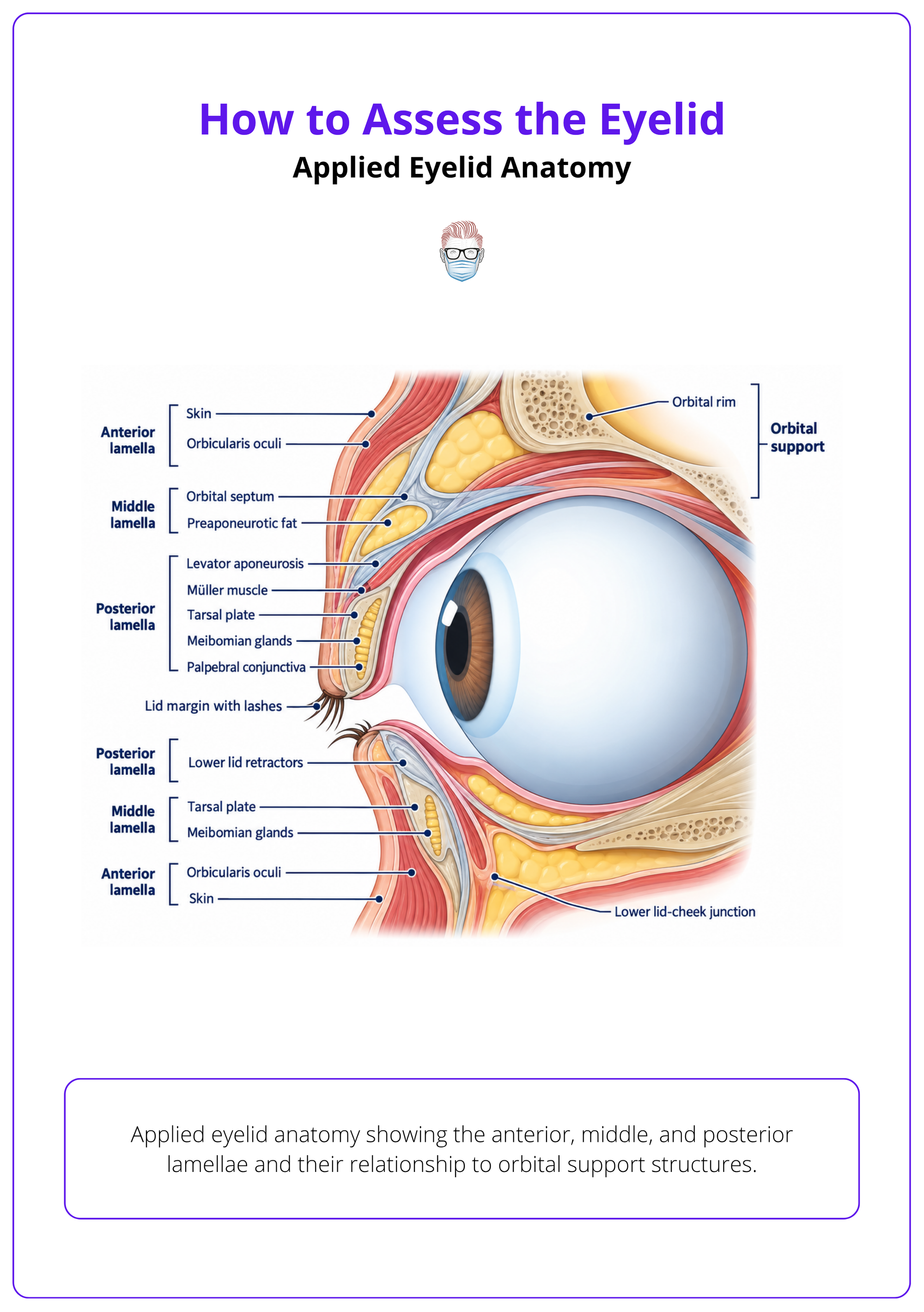
Organise the examination by anterior lamella, middle lamella, posterior lamella, and orbital support structures.
- Anterior lamella: skin and orbicularis determine redundancy, scarring, lash rotation, and closure.
- Middle lamella: septum and preaponeurotic fat affect fullness, retraction, and surgical planes.
- Posterior lamella: tarsus, conjunctiva, retractors, and canthal tendons determine margin stability.
- Lower lid planning: distinguish anterior lamellar problems from septal, retractor, tarsoconjunctival, and lid-cheek findings 1.
A structured eyelid examination can be organised by lamellar planes from skin through orbicularis, septum, fat, retractors, and tarsoconjunctiva 2.
Margin And Canthi
Examine the lid margin and canthal tendons because small abnormalities here cause major functional symptoms.
- Lid margin: record contour, notching, thickening, ulceration, lash loss, and lash misdirection.
- Tarsal plate: assess stability and meibomian gland region; MGD assessment belongs at the tarsal plate and lid margin 3.
- Canthi: check medial and lateral tendon position, rounding, laxity, and canthal tilt.
- Lacrimal drainage: inspect puncta, canaliculi, lacrimal sac region, and medial canthal scars.
Canalicular obstruction must be localised because management is difficult and depends on the obstruction site 4.
Neuromuscular Protection
Eyelid position only matters clinically when linked to opening, closure, sensation, and corneal safety.
- Levator complex: levator aponeurosis and Müller muscle determine upper lid height and crease.
- Orbicularis: facial nerve function determines blink, forced closure, and exposure risk.
- Corneal protection: lid-globe apposition, blink, Bell phenomenon, tear film, and sensation protect the cornea.
- Danger points: avoid missing facial palsy, trigeminal sensory loss, canalicular injury, or exposed cornea.
Document lid shape, contour, height, and mobility because these features determine ocular protection, function, and appearance 2.
Lamellar Eyelid Examination
Examine the eyelid by anterior lamella, middle lamella, posterior lamella, and orbital support so that visible signs map to the layer causing the deformity.
Use a clinical photograph or simple sagittal illustration to show these layers before discussing abnormal findings. This helps separate skin redundancy, septal fullness, tarsal instability, conjunctival disease, and canthal malposition during assessment 12.
- Anterior lamella: skin and orbicularis determine dermatochalasis, scarring, lash rotation, lagophthalmos, and blink strength.
- Middle lamella: septum and preaponeurotic fat influence fullness, hollowing, retraction, and safe surgical planes.
- Posterior lamella: tarsus, conjunctiva, retractors, and canthal tendons determine lid margin position, stability, and fornix health.
- Orbital support: rim, globe position, brow, and cheek support affect apparent ptosis, ectropion risk, and eyelid-cheek contour.
History and Symptoms
Focus the history on onset, fluctuation, pain, vision, diplopia, exposure symptoms, tearing, malignancy red flags, systemic disease, medication risk, and the patient’s functional or aesthetic complaint.
The history separates benign age-related eyelid change from neurological, inflammatory, orbital, lacrimal, traumatic, and malignant disease. It also tells you whether treatment is safe today.
Onset And Course
Sudden, progressive, fluctuating, and recurrent symptoms point to different diagnostic pathways.
- Sudden onset: consider trauma, infection, vascular, neurological, or acute orbital disease.
- Progressive change: ask for old photographs to distinguish involutional ptosis from new pathology.
- Fluctuation: ask about fatigability, diurnal variation, diplopia, and bulbar weakness.
- Recurrence: previous chalazion, hordeolum, surgery, filler, trauma, or cancer treatment changes risk.
For ptosis, record prior photographs, onset, associated symptoms, pupils, and extraocular movements because these help identify serious neurological or systemic causes 5.
Neurological Symptoms
Ptosis with diplopia, pupil change, headache, or systemic neurological symptoms is not cosmetic until proven otherwise.
- Diplopia: clarify binocular versus monocular, direction of gaze, variability, and associated ptosis.
- Pupil symptoms: ask about anisocoria, new miosis, mydriasis, pain, and facial sweating change.
- Myasthenia symptoms: fatigable ptosis, variable diplopia, dysphagia, dysarthria, or limb fatigue.
- Emergency symptoms: severe headache, visual loss, ophthalmoplegia, altered consciousness, or pituitary symptoms raise concern for pituitary apoplexy 6.
A suspected third nerve pattern with ptosis or diplopia should move the patient into an urgent investigation pathway rather than routine eyelid planning 7.
Ocular Surface Symptoms
Dryness, burning, foreign-body sensation, and fluctuating vision may be eyelid-driven rather than globe-driven.
- Dry eye burden: ask chronicity, artificial tear use, contact lens tolerance, screen symptoms, and nocturnal exposure.
- MGD symptoms: ocular irritation and fluctuating vision should raise suspicion for meibomian gland dysfunction 3.
- Symptom scoring: OSDI can document symptom burden when dry eye is suspected 8.
- Associated pattern: ask about sicca symptoms, autoimmune disease, rosacea, rheumatoid arthritis, and smoking.
Dry eye assessment should include eyelid-margin and meibomian gland evaluation because MGD can drive evaporative dry eye 9.
Infection And Inflammation
Pain, redness, swelling, discharge, and skin change define urgency before diagnosis.
- Swollen red lid: document onset, pain, focal mass versus diffuse swelling, vision change, and ophthalmoplegia 10.
- Hordeolum pattern: acute painful red swollen lump supports hordeolum assessment 11.
- Blepharitis symptoms: ask about itching, burning, foreign-body sensation, redness, and cylindrical dandruff 12.
- Spread: ask about fever, cellulitis, sinus symptoms, immunosuppression, and worsening despite treatment.
Lacrimal And Systemic Risk
Tearing, medication risk, and systemic disease can change both diagnosis and operative safety.
- Epiphora: ask unilateral versus bilateral, constant versus intermittent, discharge, blood-stained tears, and sac swelling.
- Malignancy warning: persistent or isolated epiphora needs careful assessment because lacrimal drainage carcinoma may present after delayed symptoms 13.
- Treatment risk: document anticoagulants, antiplatelets, steroids, immunosuppression, isotretinoin history, and drug allergies.
- Thyroid disease: ask about eye prominence, lid retraction, diplopia, exposure, smoking, and thyroid control.
Epiphora should not be assumed benign because lacrimal drainage system carcinoma can resemble inflammatory obstruction 13.
Inspection and Static Examination
Before touching the eyelid, inspect facial symmetry, brow position, lid height, lid margin, lashes, canthi, scleral show, globe position, skin quality, scars, swelling, lesions, and conjunctival inflammation.
Static inspection sets the diagnosis before measurement. Examine the patient sitting upright, looking straight ahead, with the brow relaxed and spectacles removed.
Facial Symmetry
Inspect the whole periocular unit because brow, cheek, malar, and temporal descent can mimic eyelid disease.
- Brow position: distinguish brow ptosis from upper lid dermatochalasis.
- Periocular ageing: record hooding, lateral temporo-orbital sagging, orbicularis descent, lid-cheek depth, and festoons 14.
- Facial palsy: look for brow descent, widened fissure, poor blink, lower lid descent, and oral asymmetry.
- Globe prominence: compare proptosis, enophthalmos, hypoglobus, and asymmetry.
Assess eyebrow position, orbicularis status, lateral temporal sagging, lid-cheek junction, and malar change together rather than judging the eyelid alone 14.
Eyelid Position
Describe lid position objectively before deciding whether the problem is ptosis, retraction, malposition, or skin excess.
- Upper lid: look for low margin, high crease, asymmetry, lash ptosis, and frontalis recruitment.
- Lower lid: inspect ectropion, entropion, scleral show, retraction, rounding, and lid-globe apposition.
- Canthal tilt: compare medial and lateral canthi, noting rounding or lateral descent.
- Resting closure: identify lagophthalmos before asking for forced closure.
Static eyelid inspection should record position and symmetry using standardised measurements rather than subjective description alone 5.
Margin And Lashes
The lid margin is a diagnostic surface for inflammation, scarring, malignancy, and lash-related corneal risk.
- MGD signs: record plugging, capping, foamy tear, telangiectasia, and gland expressibility.
- Blepharitis signs: assess redness, duct condition, crusting, lash adhesions, telangiectasia, and surrounding irritation 15.
- Trichiasis: upper-lid lash misdirection with conjunctival scarring can threaten sight 16.
- Demodex: cylindrical eyelash collarettes on slit-lamp inspection support ocular Demodex assessment 17.
Lesions And Skin
Localise eyelid lesions by margin involvement, skin change, lash loss, ulceration, and fixation.
- Margin lesions: examine closely when SCC is suspected because eyelid SCC commonly involves the margin 18.
- Mid-lid mass: a central intratarsal mass often supports chalazion in the differential 10.
- Lid-margin mass: a well-defined painful margin lesion supports hordeolum or stye 10.
- Skin quality: note scars, radiation change, actinic damage, inflammation, oedema, and previous incisions.
Photograph primary gaze, upgaze, downgaze, gentle closure, and any focal lesion when comparison or treatment planning is likely.
Functional Eyelid Measurements
Measure MRD1, MRD2, palpebral fissure height, levator function, crease height, lagophthalmos, orbicularis strength, Bell phenomenon, and lid laxity to define severity and operative risk.
Functional measurement turns inspection into a defensible assessment. Measure both sides, control frontalis activity, and record conditions that may alter lid height.
Margin Reflex Distance
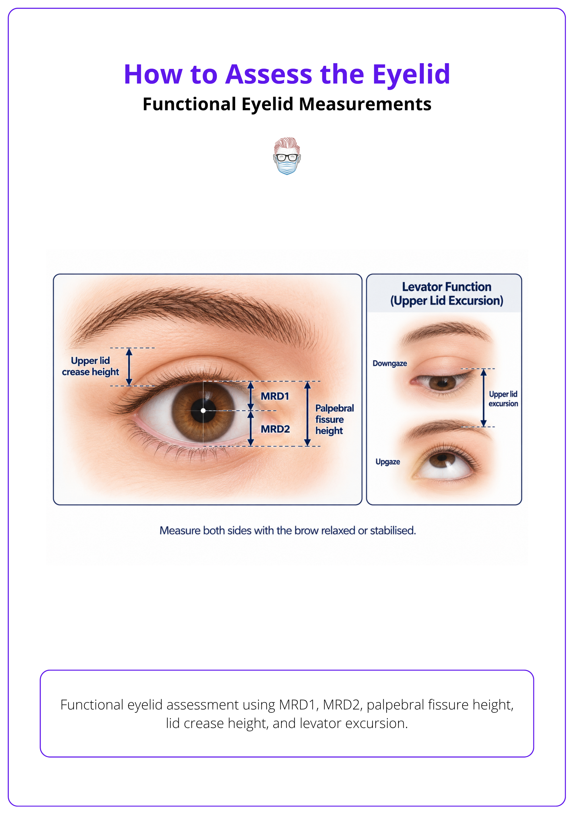
Use the corneal light reflex as the reference point for upper and lower lid position.
- MRD1: distance from corneal reflex to upper lid margin; primary objective ptosis measurement.
- MRD2: distance from corneal reflex to lower lid margin; helps define lower lid retraction or elevation.
- Fissure height: vertical aperture between upper and lower lid margins.
- Drug effect: recent oxymetazoline 0.1% can alter MRD1 and should be documented 19.
Functional eyelid assessment should include MRD1, MRD2, and palpebral fissure height when evaluating eyelid opening or ptosis-related appearance 19.
| Measurement | What It Defines | Practical Use |
|---|---|---|
| MRD1 | Upper lid height | Ptosis severity |
| MRD2 | Lower lid position | Retraction or scleral show |
| Palpebral fissure | Lid aperture | Global asymmetry |
| Levator function | Lid excursion | Procedure planning |
| Lagophthalmos | Closure deficit | Corneal exposure risk |
| Distraction test | Horizontal laxity | Canthal support need |
Levator Function
Measure lid excursion with the brow immobilised so frontalis compensation does not hide poor levator function.
- Technique: stabilise the brow and measure excursion from downgaze to upgaze.
- Crease height: high crease suggests aponeurotic dehiscence; absent crease may suggest congenital ptosis.
- Superior rectus: check upgaze because poor elevation affects ptosis planning.
- Synkinesis: look for jaw-winking, aberrant regeneration, or paradoxical lid movement.
Before ptosis surgery, measure severity, levator function, superior rectus action, Bell phenomenon, lagophthalmos, and synkinetic movement 20.
Eyelid Closure
Closure testing defines corneal risk and determines whether eyelid elevation or skin excision is safe.
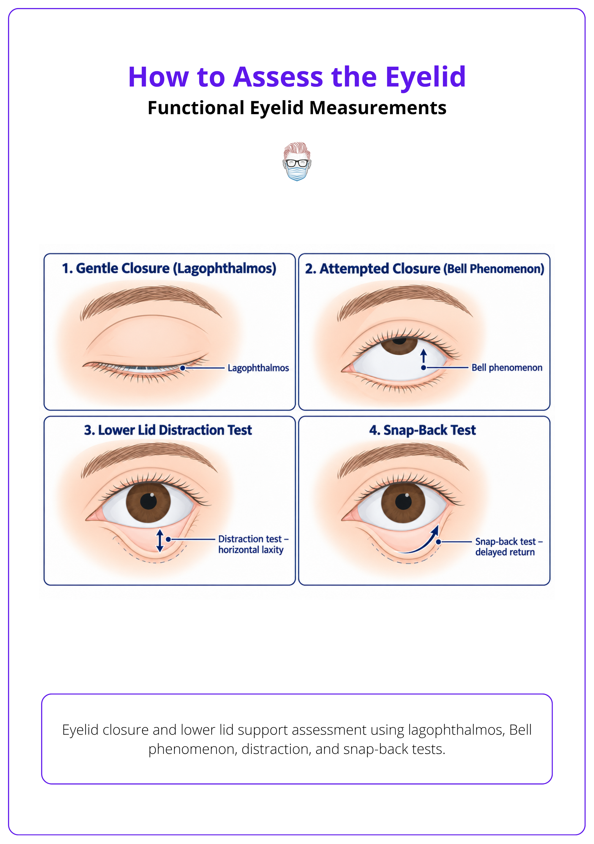
- Gentle closure: look for lagophthalmos without forced squeezing.
- Forced closure: assess orbicularis power and facial nerve function.
- Bell phenomenon: document upward globe rotation during attempted closure.
- Exposure risk: combine closure, tear film, corneal sensation, and lower lid support.
Follow-up after eyelid-opening procedures should reassess lagophthalmos, corneal exposure, position, excursion, synkinesis, and symmetry 20.
Lid Laxity
Laxity testing separates horizontal instability from vertical malposition and muscle weakness.
- Distraction test: pull lid away from globe and compare side-to-side.
- Snap-back test: delayed return suggests lower lid laxity or orbicularis weakness.
- Canthal tendons: assess medial and lateral tendon laxity separately.
- Separate domains: eyelid laxity does not automatically mean reduced fissure height or poor levator function 21.
Assess eyelid position using laxity, dermatochalasis, interpalpebral distance, and levator function rather than a single visual impression 21.
Ocular Surface and Lacrimal Assessment
Check visual acuity, corneal staining, tear film, dry eye features, punctal position, lacrimal sac findings, and drainage patency before planning eyelid intervention.
No eyelid plan is safe until the cornea and tear system have been assessed. A lid that looks acceptable can still be dangerous if closure, sensation, tear film, or drainage is poor.
Visual Screening
Screen vision and corneal safety early because eyelid treatment can worsen exposure.
- Visual acuity: document baseline acuity and any acute change.
- Cornea: inspect for exposure, epithelial defect, ulceration, opacity, or reduced sensation.
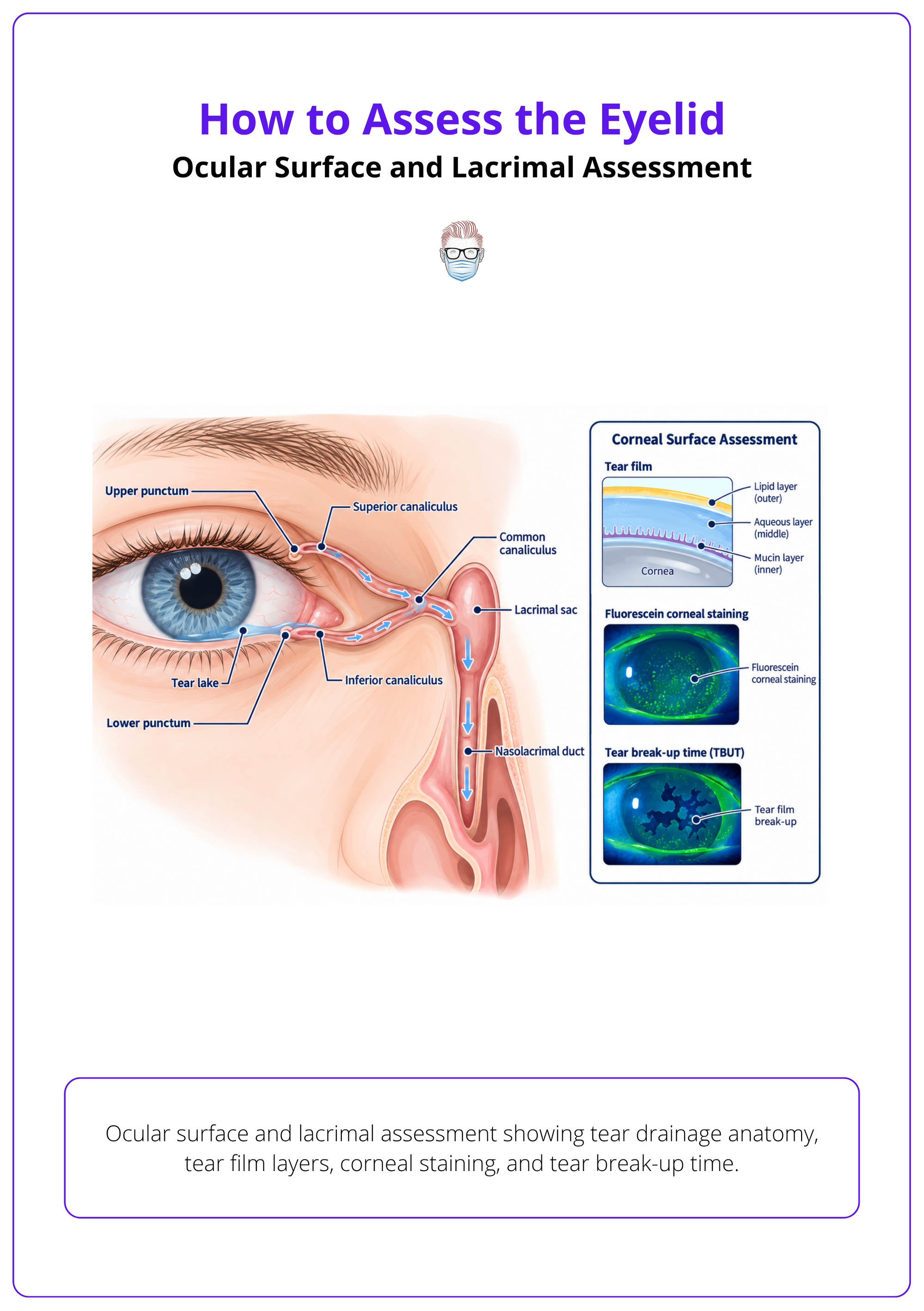
- Fluorescein: stain when exposure, irritation, trichiasis, entropion, or dry eye is suspected.
- Slit lamp: involve ophthalmology when corneal compromise or diagnostic uncertainty exists.
Dry eye evaluation should not depend on a single test; symptoms and ocular surface or tear-film findings must be interpreted together.
Tear Film
Tear symptoms need objective surface findings before attributing them to eyelid position alone.
- TBUT: record fluorescein tear break-up time in seconds when assessing tear-film instability 3.
- Schirmer: record wetting in mm over 5 minutes when tear production assessment is needed 8.
- Staining: higher corneal and conjunctival staining indicates greater ocular surface abnormality 8.
- MGD: include lid-margin and meibomian gland evaluation in dry eye assessment.
Ocular surface assessment may include OSDI, TBUT, Schirmer testing, corneal fluorescein staining, lissamine green staining, tear osmolarity, and MGD grading 8.
Lacrimal Drainage
Epiphora assessment should localise punctal, canalicular, sac, duct, and pump problems.
- Puncta: inspect position, stenosis, eversion, obstruction, and apposition to the globe.
- Lacrimal sac: palpate for swelling, tenderness, reflux, mass, or discharge.
- Regurgitation: pressure over the sac may reveal mucous, pus, or blood-stained reflux.
- Syringing: use when obstruction localisation will change referral or treatment.
Define whether stenosis or obstruction is partial or complete because lacrimal terminology is not uniform 4.
Pattern Recognition and Differential Diagnosis
Organise findings into position disorders, inflammatory or infective patterns, lacrimal disease, neurological causes, orbital disease, trauma, and benign versus malignant lesions.
Pattern recognition prevents the common error: treating dermatochalasis, ptosis, brow descent, and eyelid malposition as the same problem. Name the dominant pattern before proposing intervention.
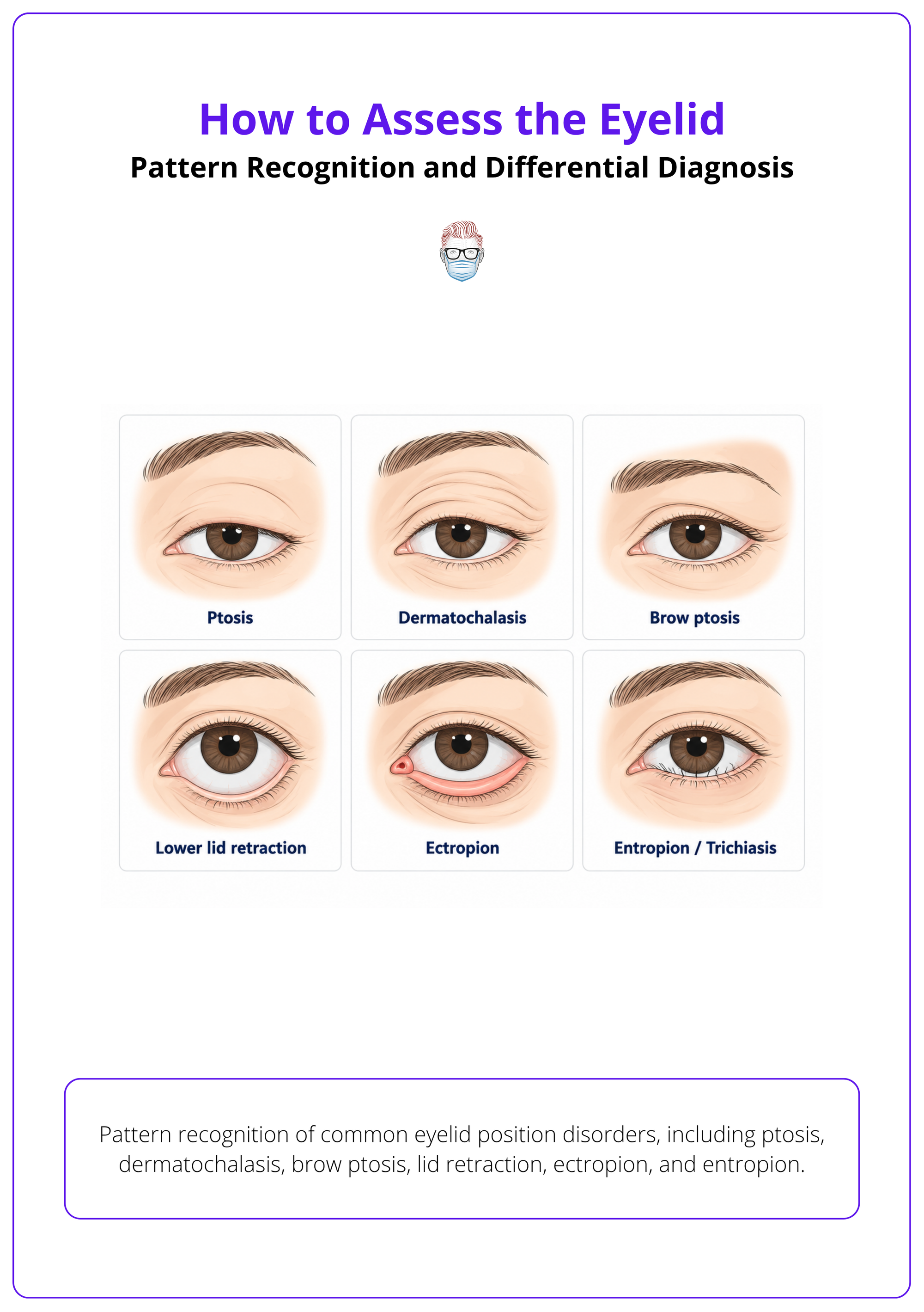
Position Disorders
Separate true ptosis from pseudoptosis and malposition before choosing treatment.
- Ptosis: low upper lid margin; assess levator, pupil, motility, and variability.
- Dermatochalasis: redundant skin with relatively preserved lid margin position.
- Brow ptosis: low brow causing secondary hooding, especially laterally.
- Retraction: high upper lid or low lower lid, often with thyroid, scarring, or orbital disease.
The eyelid assessment should distinguish true ptosis from pseudoptosis and masquerade conditions before treatment is selected 5.
| Dominant Finding | Likely Pattern | Next Assessment Move |
|---|---|---|
| Low upper margin | Ptosis | Levator, pupils, motility |
| Skin hooding | Dermatochalasis | Brow position, field symptoms |
| Low brow | Brow ptosis | Frontalis use, asymmetry |
| Lower scleral show | Retraction | Thyroid and orbital signs |
| Out-turned lid | Ectropion | Laxity and punctal position |
| In-turned lid | Entropion | Lashes and corneal staining |
Red Eyelid Patterns
Pain, vision change, ophthalmoplegia, vesicles, plaques, and swelling distribution define urgency.
- Preseptal cellulitis: lid swelling without orbital signs.
- Orbital cellulitis: reduced eye movement or vision change raises concern for orbital rather than preseptal disease 10.
- Inflammatory mimics: vesicles, irritation, raised plaques, and malignant changes help distinguish zoster, contact dermatitis, atopic dermatitis, and skin cancer 10.
- Cellulitis distinction: swollen red eyelid assessment must separate preseptal from orbital cellulitis because urgency differs 10.
Lesion Patterns
Eyelid lesions require suspicion when they distort architecture, recur, bleed, ulcerate, or cause lash loss.
- Benign lumps: chalazion is usually intratarsal; hordeolum is often painful and margin-related.
- Malignancy: suspect with ulceration, induration, lash loss, bleeding, pearly edge, pigmentation, or recurrence.
- Adnexal tumours: rare adnexal carcinoma can be misdiagnosed, so atypical lesions deserve pathology 22.
- Floppy eyelid: consider obstructive sleep apnoea when floppy eyelid syndrome is recognised 23.
When eyelid anomalies occur with ear anomalies, midface hypoplasia, and micrognathia, consider facial dysostosis syndromes 24.
Investigations and Referral Triggers
Investigate or refer urgently for corneal compromise, vision change, orbital signs, acute neurological ptosis, suspected malignancy, canalicular injury, severe infection, or unclear lacrimal obstruction.
Most eyelid assessments need no imaging. Tests are justified when they change safety, diagnosis, urgency, or procedure selection.
Ophthalmic Testing
Use eye tests when function, corneal safety, or diagnosis cannot be judged by inspection alone.
- Visual fields: request when ptosis or dermatochalasis causes superior field symptoms or functional surgery is considered.
- Slit lamp: seek assessment for corneal staining, ulceration, intraocular inflammation, trichiasis injury, or uncertain surface disease.
- Myasthenia: investigate fluctuating ptosis, variable diplopia, fatigability, or misalignment not fitting a cranial nerve pattern.
- Thyroid disease: arrange thyroid tests and ophthalmology input when retraction, proptosis, diplopia, or exposure suggests thyroid eye disease.
If ocular alignment symptoms do not match a cranial nerve palsy pattern, ocular myasthenia should be considered 7.
Neuroimaging
Acute neurological eyelid signs require escalation before eyelid treatment.
- Third nerve palsy: ptosis with diplopia in a third nerve pattern should trigger urgent brain CTA to exclude aneurysm 7.
- Multiple cranial nerves: urgent neuroimaging should focus on the cavernous sinus and superior orbital fissure 7.
- Brainstem symptoms: recent binocular diplopia with brainstem symptoms warrants urgent brain MRI 7.
- Pituitary apoplexy: MRI is preferred when ocular motor signs suggest pituitary apoplexy 6.
Horner syndrome should not be excluded simply because ptosis, miosis, and anhidrosis are not all present 25.
Biopsy And Referral
Refer when tissue diagnosis, drainage reconstruction, orbital care, or emergency eye protection is needed.
- Suspicious lesions: biopsy or oculoplastic referral for ulceration, lash loss, bleeding, recurrence, fixation, pigmentation, or architectural distortion.
- Pathology review: suspicious adnexal lesions need careful pathology review because misdiagnosis has been reported 22.
- Canalicular laceration: refer for repair planning; repair with silicone intubation has reported high success 4.
- Lacrimal malignancy: persistent obstructive lacrimal symptoms need timely investigation because delayed diagnosis can lead to advanced disease and nodal spread 13.
Patients with floppy eyelid syndrome or other OSA-associated ocular findings should be screened for obstructive sleep apnoea and referred when appropriate 23.
Documentation and Assessment Summary
Record laterality, photographs, drawings, measurements, ocular surface findings, red flags, diagnosis, severity, functional impact, treatment risks, and a clear plan for observation, referral, or intervention.
Good eyelid documentation allows comparison over time and protects the patient from poorly justified intervention. It should be structured enough that another clinician can understand the diagnosis without re-examining from scratch.
Structured Charting
Document what was measured, what was seen, and what risks change management.
- Laterality: record side, symmetry, brow position, lid height, margin findings, and canthal support.
- Measurements: include MRD1, MRD2, fissure height, levator function, lagophthalmos, and laxity tests.
- Surface findings: document vision, cornea, fluorescein staining, tear film, puncta, and lacrimal sac findings.
- Urgency data: onset, pain, focal mass versus diffuse swelling, vision change, and ophthalmoplegia guide diagnosis and urgency 10.
Documentation should be meticulous because structured eyelid assessment improves recognition and outcomes in acquired ptosis 5.
Assessment Impression
Finish with a working diagnosis, severity statement, functional impact, and next action.
- Photographs: standardise primary gaze, oblique views, closure, upgaze, downgaze, and lesion close-ups.
- Tumour summary: for suspicious eyelid tumours, capture demographics, presentation, diagnosis, treatment plan, and follow-up 22.
- Blepharoplasty planning: document patient-reported outcomes, scars, eyebrow height, tarsal platform show, and aesthetic assessment 26.
- Final plan: state observation, lubrication, investigation, ophthalmology referral, biopsy, surgery, or emergency escalation.
References
[1] Mojallal et al.. Anatomy of lower eyelid and eyelid-cheek junction.. Annales de chirurgie plastique et esthetique. 2017. doi:10.1016/j.anplas.2017.09.007
[2] Dailey et al.. Eyelid anatomy.. The Journal of dermatologic surgery and oncology. 1992. doi:10.1111/j.1524-4725.1992.tb02779.x
[3] Chen et al.. Which treatment works better for Meibomian Gland Dysfunction: LipiFlow or intense pulsed light? A systematic review and network meta-analysis.. Photodiagnosis and photodynamic therapy. 2025. doi:10.1016/j.pdpdt.2025.104630
[4] Ali et al.. Human Lacrimal Drainage System Reconstruction, Recanalization, and Regeneration.. Current eye research. 2020. doi:10.1080/02713683.2019.1580376
[5] Kirschen et al.. Topical Review: An Update of Diagnostic and Management Algorithms for Acquired Blepharoptosis. Optometry and Vision Science. 2022. doi:10.1097/OPX.0000000000001868
[6] Murad-Kejbou et al.. Pituitary apoplexy: evaluation, management, and prognosis.. Current opinion in ophthalmology. 2009. doi:10.1097/ICU.0b013e3283319061
[7] Margolin. Approach to patient with diplopia.. Journal of the neurological sciences. 2021. doi:10.1016/j.jns.2020.117055
[8] Yu et al.. Systemic Conditions Associated with Severity of Dry Eye Signs and Symptoms in the Dry Eye Assessment and Management Study.. Ophthalmology. 2021. doi:10.1016/j.ophtha.2021.03.030
[9] McCann et al.. Prevalence and Incidence of Dry Eye and Meibomian Gland Dysfunction in the United States: A Systematic Review and Meta-analysis.. JAMA ophthalmology. 2022. doi:10.1001/jamaophthalmol.2022.4394
[10] Carlisle et al.. Differential Diagnosis of the Swollen Red Eyelid.. American family physician. 2015. PMID:26176369
[11] Lindsley et al.. Non-surgical interventions for acute internal hordeolum.. The Cochrane database of systematic reviews. 2017. doi:10.1002/14651858.CD007742.pub4
[12] Kairey et al.. Efficacy and safety of Melaleuca alternifolia (tea tree) oil for human health-A systematic review of randomized controlled trials.. Frontiers in pharmacology. 2023. doi:10.3389/fphar.2023.1116077
[13] Ramberg et al.. Carcinomas of the lacrimal drainage system.. Survey of ophthalmology. 2020. doi:10.1016/j.survophthal.2020.04.001
[14] Pascali et al.. The Temporal Subcutaneous Brow Lift with Orbicularis Oculi Muscle Elastic Flap: Technical Considerations, Systematic Review, and Terminology Standardization.. Facial plastic surgery : FPS. 2023. doi:10.1055/a-1953-2304
[15] Siegel et al.. BlephEx-treatment for blepharitis: a prospective randomized placebo-controlled trial.. BMC ophthalmology. 2024. doi:10.1186/s12886-024-03765-3
[16] Yorston et al.. Interventions for trachoma trichiasis.. The Cochrane database of systematic reviews. 2006. doi:10.1002/14651858.CD004008.pub2
[17] Zhang et al.. Ocular Demodex: a systematic review of the clinical literature.. Ophthalmic & physiological optics : the journal of the British College of Ophthalmic Opticians (Optometrists). 2020. doi:10.1111/opo.12691
[18] Watson et al.. Adnexal squamous cell carcinoma: incidence of eyelid margin involvement.. Orbit (Amsterdam, Netherlands). 2023. doi:10.1080/01676830.2022.2092156
[19] Shoji et al.. The effects of topical oxymetazoline on eyelid position, eye redness, and patient-reported eye appearance: A randomized controlled trial.. Journal of plastic, reconstructive & aesthetic surgery : JPRAS. 2023. doi:10.1016/j.bjps.2023.02.006
[20] Ibrahim. Use of the levator muscle as a frontalis sling.. Ophthalmic plastic and reconstructive surgery. 2008. doi:10.1097/IOP.0b013e3181379e20
[21] Karaca et al.. Evaluation of Periorbital Tissues in Obstructive Sleep Apnea Syndrome.. Turkish journal of ophthalmology. 2021. doi:10.4274/tjo.galenos.2020.35033
[22] Froehlich et al.. Endocrine mucin-producing sweat gland carcinoma: a systematic review and meta-analysis.. The Journal of dermatological treatment. 2022. doi:10.1080/09546634.2021.1937479
[23] Bulloch et al.. Ocular manifestations of obstructive sleep apnea: a systematic review and meta-analysis.. Graefe's archive for clinical and experimental ophthalmology = Albrecht von Graefes Archiv fur klinische und experimentelle Ophthalmologie. 2024. doi:10.1007/s00417-023-06103-3
[24] van et al.. Unveiling the Phenotypic Spectrum of Miller Syndrome: A Systematic Review\.. The Journal of craniofacial surgery. 2025. doi:10.1097/SCS.0000000000011501
[25] Davagnanam et al.. Adult Horner's syndrome: a combined clinical, pharmacological, and imaging algorithm.. Eye (London, England). 2013. doi:10.1038/eye.2012.281
[26] Hollander et al.. Aesthetic outcomes of upper eyelid blepharoplasty: a systematic review\.. International journal of oral and maxillofacial surgery. 2020. doi:10.1016/j.ijom.2019.10.014
