


Summary Card
Assessment Goals
Fat distribution assessment should confirm excess adiposity, define regional and visceral patterns, document contour imbalance or donor adequacy, and identify patients needing risk modification or referral.
Clinical History
History should define weight stability, metabolic and endocrine context, prior operations, pregnancy or bariatric history, oedema symptoms, medication effects, and the patient’s actual contour or grafting goal.
Physical Examination
Examine fat distribution systematically by region, confirming pinchable subcutaneous fat, skin quality, fascial integrity, oedema, asymmetry, scars, hernias, and reproducible documentation.
Measurement and Classification
Use BMI for overall size, waist-based measures for central adiposity, anthropometry or body composition tools for compartment pattern, and named classifications such as android, gynoid, generalised, regional, or abnormal distribution.
Investigations and Imaging
Use imaging or tests selectively when clinical assessment cannot distinguish visceral fat, oedema, hernia, body composition, endocrine disease, or donor-site safety.
Pattern Recognition and Differential Diagnosis
Abnormal fat distribution is recognised by disproportion, pain, oedema, metabolic context, endocrine features, medication exposure, syndromic clues, or contour change after surgery.
Treatment Planning Implications
Fat distribution assessment directs weight optimisation, liposuction, excisional contouring, donor-site selection, staged surgery, hernia repair, metabolic referral, endocrine review, and risk counselling.
Assessment Goals
Fat distribution assessment should confirm excess adiposity, define regional and visceral patterns, document contour imbalance or donor adequacy, and identify patients needing risk modification or referral.
Assessing fat distribution is not the same as recording weight. The clinical task is to decide whether the patient has a contour problem, a metabolic-risk pattern, an abnormal fat disorder, or a surgical planning issue.
Clinical Purpose
Use fat distribution assessment to answer the decision that BMI cannot answer.
- Regional adiposity: identify localised subcutaneous fat suitable for contouring or donor harvest.
- Central adiposity: supplement BMI with waist circumference, waist-to-height ratio, or related measures when central risk matters 1.
- Body composition: recognise that BMI can miss clinically relevant differences in fat, muscle, and bone composition 2.
- Surgical planning: include fat-distribution assessment when body composition may influence operative planning or risk counselling 3.
Planning Questions
Convert the examination into a treatment, referral, or deferral decision.
- Aesthetic contouring: separate pinchable subcutaneous fat from visceral fullness, skin excess, or fascial laxity.
- Reconstructive planning: assess whether donor fat is adequate, safe, and proportionate to recipient requirements.
- Risk stratification: consider regional adipose patterns when cardiometabolic risk is part of the clinical question 4.
- Diagnostic uncertainty: integrate history, examination, and tests when oedema or limb enlargement may mimic adiposity 5.
Clinical History
History should define weight stability, metabolic and endocrine context, prior operations, pregnancy or bariatric history, oedema symptoms, medication effects, and the patient’s actual contour or grafting goal.
The history changes how fat distribution is interpreted. A stable saddlebag deformity, central visceral obesity, post-bariatric skin redundancy, and painful limb enlargement are different clinical problems.
Weight History
Establish whether the current pattern is stable, changing, or part of active weight disease.
- Weight trajectory: document maximum weight, lowest adult weight, recent change, and duration of stability.
- Bariatric history: record procedure type, date, complications, nutritional status, and weight plateau before body contouring.
- Diet pattern: distinguish diet quality because unhealthy plant-based dietary patterns have been linked with abdominal obesity risk 6.
- Recovery states: previous anorexia nervosa or nutritional rehabilitation should prompt assessment for regional fat redistribution rather than assuming uniform low fat 7.
Medical History
Identify diseases and drugs that can create misleading or high-risk fat patterns.
- Metabolic risk: screen for diabetes, hypertension, dyslipidaemia, NAFLD, sleep apnoea, and mechanical symptoms.
- Complication burden: history should screen for the presence and severity of weight-related complications and be paired with laboratory review 1.
- Mood context: if depression risk is part of the referral, BMI and waist-to-height ratio may be insufficient without considering visceral adiposity 8.
- Medication effects: ask about corticosteroids, antiretrovirals, insulin, antipsychotics, hormonal therapy, and weight-loss drugs.
Symptom History
Pain, swelling, bruising, and functional limitation shift the assessment from contour analysis to diagnosis.
- Lipedema clue: painful, disproportionate limb fat in a female patient should raise suspicion for lipedema 9.
- Oedema clue: unilateral lower-limb swelling history should include cardiac, hepatic, renal, trauma, malignancy, radiation, and surgical factors 5.
- Functional impact: ask about heaviness, mobility restriction, clothing difficulty, exercise limitation, and recurrent cellulitis.
- Patient goals: body-contouring assessment should include open discussion of the patient’s goals and concerns 10.
Physical Examination
Examine fat distribution systematically by region, confirming pinchable subcutaneous fat, skin quality, fascial integrity, oedema, asymmetry, scars, hernias, and reproducible documentation.
Examination should distinguish fat from skin, muscle, oedema, hernia, and posture. The patient should be assessed standing where possible, with adequate exposure and chaperone support.
Inspection
Inspect the whole aesthetic or donor unit before focusing on the patient’s complaint.
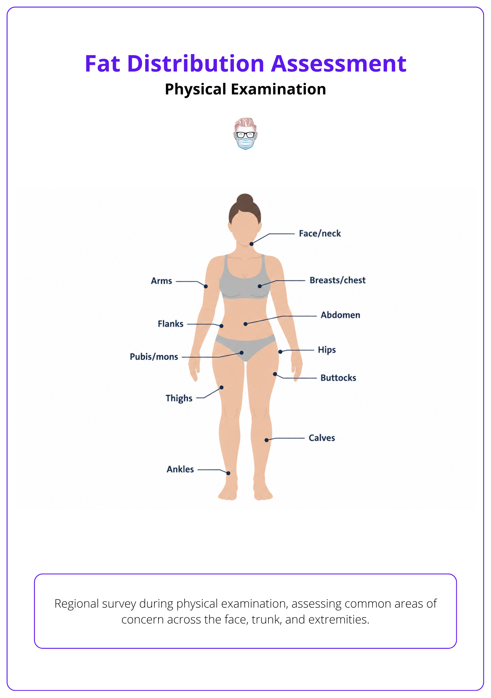
- Regional survey: assess face, neck, trunk, flanks, arms, breasts, abdomen, pubis, buttocks, hips, thighs, calves, and ankles.
- Pattern symmetry: document unilateral, bilateral, upper-body, lower-body, central, peripheral, or segmental distribution.
- Surface contour: look for bulges, hollows, step-offs, skin tethering, cellulite, rolls, and post-liposuction irregularity.
- Posture effect: reassess abdominal and flank contour with relaxed posture, lordosis correction, and muscle activation.
Palpation
Confirm whether the contour is soft subcutaneous fat, firm fibrosis, fluid, hernia, or fascial laxity.
- Pinch assessment: estimate subcutaneous thickness, tissue mobility, tenderness, and whether liposuction can safely treat the layer.
- Skin quality: record elasticity, striae, dermal thinning, redundancy, scars, pigmentation, and intertrigo.
- Oedema signs: assess pitting, Stemmer sign, temperature, skin thickening, and distribution of accumulated fluid 5.
- BMI discordance: confirm whether elevated BMI reflects fat rather than lean mass, oedema, or another contributor 1.
Central Measures
Measure central adiposity reproducibly when metabolic risk or abdominal planning is relevant.
- Waist circumference: use it as an office measure of central abdominal adiposity 1.
- Technique: measure privately with a tension-controlled tape just above the anterior superior iliac spine, horizontal to the floor 1.
- PCOS caveat: waist-circumference impressions may not match MRI or CT findings in PCOS, so avoid over-reliance when decisions are high-stakes 2.
- Serial review: bodyweight, waist circumference, and BMI can be tracked during obesity-treatment follow-up 11.
Abdominal Wall
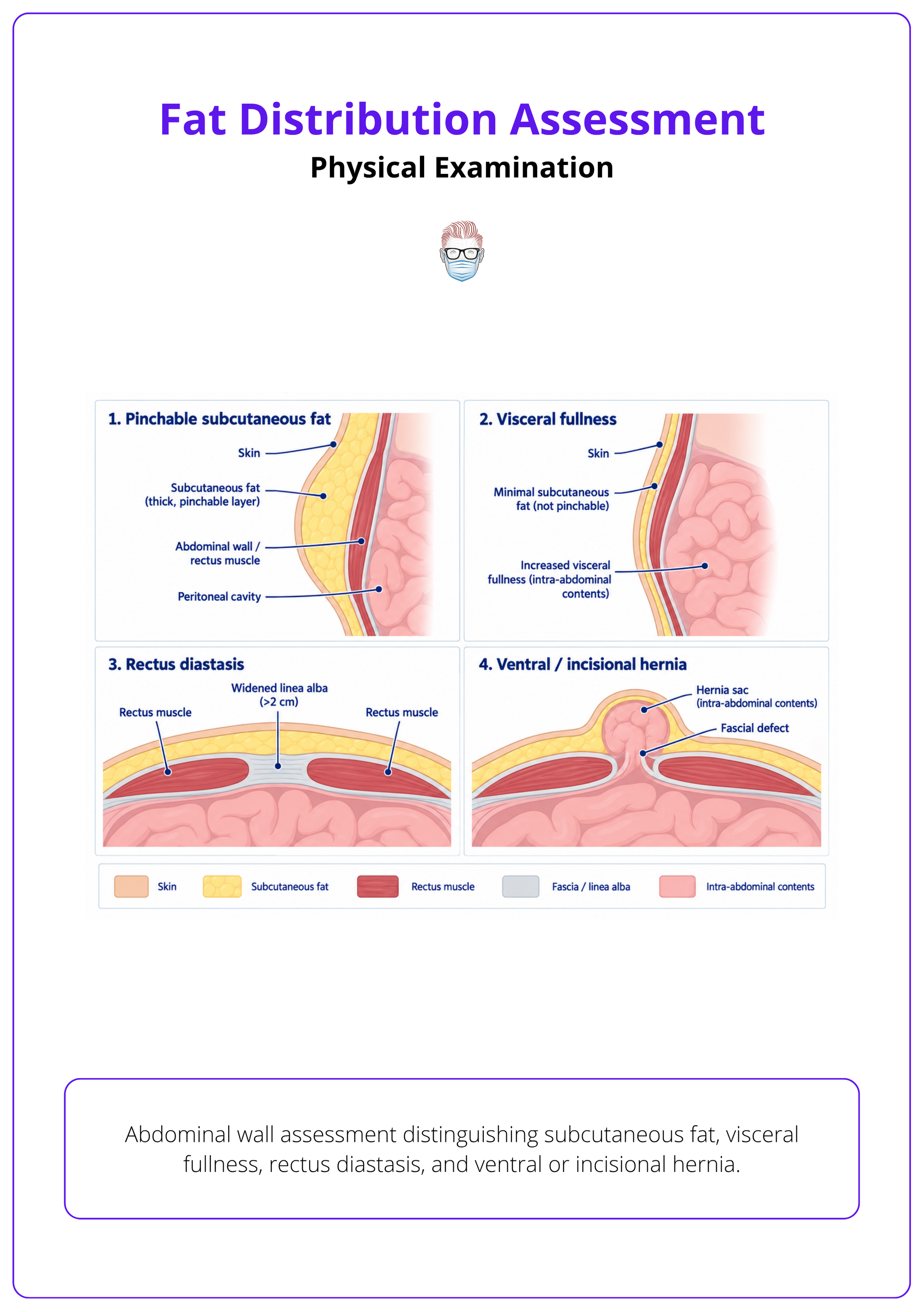
Separate adiposity from skin excess, diastasis, hernia, and intra-abdominal fullness before planning contour surgery.
- Subcutaneous fat: pinch the upper abdomen, lower abdomen, flanks, mons, and back rolls separately.
- Visceral fullness: suspect intra-abdominal fat when the abdomen remains protuberant despite limited pinchable fat.
- Fascial laxity: assess rectus diastasis, cough impulse, prior mesh, incisional hernia, and abdominal wall competence.
- Surgical scars: map Caesarean, laparotomy, stoma, drain, liposuction, and bariatric scars because they affect vascularity and access.
Documentation
Record findings in a way that another clinician can reproduce and compare.
- Photography: obtain standardised views with consistent lighting, distance, posture, and underwear position.
- Body maps: annotate fat deposits, skin excess, scars, asymmetry, hernias, tenderness, and oedema zones.
- Measurement domains: document waist circumference, waist-to-hip ratio, skinfolds, fat mass, visceral fat, and subcutaneous fat when relevant 12.
- Infant caveat: in infants, BMI should not be used as a proxy for preperitoneal abdominal fat thickness 13.
Measurement and Classification
Use BMI for overall size, waist-based measures for central adiposity, anthropometry or body composition tools for compartment pattern, and named classifications such as android, gynoid, generalised, regional, or abnormal distribution.
Measurements should support the clinical decision rather than replace examination. Record the method, landmarks, equipment, and patient position.
Anthropometry
Use simple measures to quantify the pattern seen on examination.
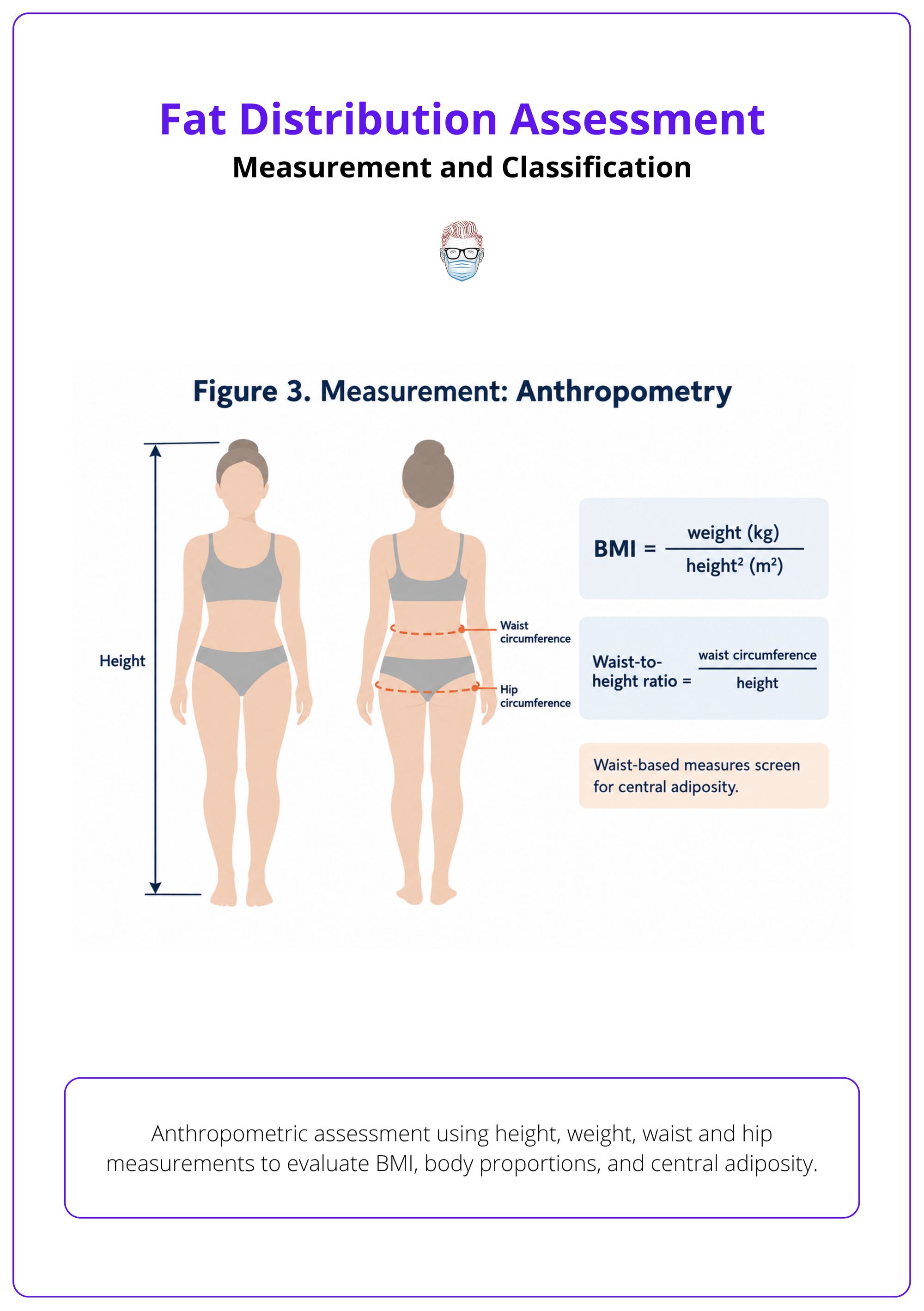
- BMI: classify overall adiposity severity, but do not treat it as a direct regional fat measurement 14.
- Waist circumference: estimate central abdominal adiposity and follow change over time.
- Hip circumference: combine with waist measurement when central versus lower-body pattern matters.
- Skinfolds: use consistent caliper sites to estimate subcutaneous thickness, especially in serial assessment.
| Measure | Best Use | Main Limitation |
|---|---|---|
| BMI | Overall adiposity category | Does not localise fat 14 |
| Waist circumference | Central adiposity screen | Technique-sensitive |
| Waist-to-hip ratio | Android versus gynoid tendency | Hip size may reflect muscle or bone |
| Skinfolds | Subcutaneous fat estimate | Operator-dependent |
| DXA or CT indices | Compartment assessment | Cost, access, radiation for CT |
Central Adiposity
Classify central fat separately because it changes metabolic counselling and referral.
- Practical screen: combine BMI confirmation with waist circumference, waist-to-height ratio, or a related central-adiposity measure 1.
- Waist-to-height rule: advise that waist circumference should remain less than half of height for pragmatic risk communication 1.
- WHtR categories: 0.4–0.49 is healthy central adiposity, 0.5–0.59 is increased risk, and 0.6 or more indicates higher risk 111.
- Anthropometric value: waist-based measures can help distinguish visceral from subcutaneous patterns and support cardiometabolic risk assessment 15.
Body Composition
Select body composition tools when the question is compartmental rather than cosmetic.
- Named domains: structure the assessment around waist circumference, waist-to-hip ratio, fat mass, fat-free mass, visceral fat, subcutaneous fat, and skinfold thickness 12.
- BMI categories: use WHO categories for overall severity: overweight 25–29.9 kg/m², class I 30–34.9, class II 35–39.9, and class III ≥40 1.
- Direct measures: excess adiposity may be classified using BMI and/or fat measures such as waist circumference, CT/DXA indices, and skinfolds 6.
- Disproportionate depots: BMI-independent fat z-scores can help identify regional depots that are out of proportion to overall body size 4.
Pattern Classification
Name the distribution pattern before deciding whether it is normal, risky, or pathological.
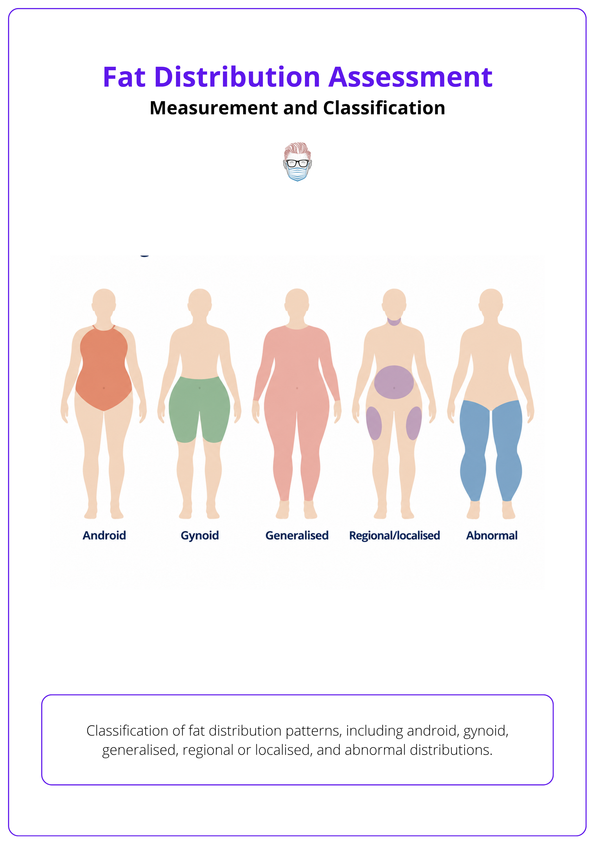
- Android pattern: upper-body trunk and abdominal predominance, often relevant to central risk assessment.
- Gynoid pattern: hips, buttocks, and thighs predominate, often with more peripheral subcutaneous distribution.
- Visceral pattern: intra-abdominal fat predominates and requires metabolic-risk consideration.
- Abnormal pattern: classify painful limb fat, lipodystrophy, oedema, post-surgical deformity, or syndromic change separately.
Investigations and Imaging
Use imaging or tests selectively when clinical assessment cannot distinguish visceral fat, oedema, hernia, body composition, endocrine disease, or donor-site safety.
Most contour assessments do not need advanced imaging. Tests add value when they answer a question that will change treatment, referral, or consent.
Cross-Sectional Imaging
Choose CT or MRI when a direct compartment measure is needed.
Body Composition Tests
Match the test to the compartment that changes management.
- DXA: useful for whole-body and regional composition estimates, but interpret cautiously when hydration of lean soft tissue may confound measurements 2.
- MRI detail: MRI can quantify visceral fat, abdominal subcutaneous fat, and liver fat when detailed regional assessment is needed 4.
- Lipedema comparison: affected thigh subcutaneous fat may differ from lower abdominal fat, so regional comparison can support disease-pattern assessment 17.
Laboratory Assessment
Order blood tests when distribution suggests systemic disease rather than isolated contour imbalance.
- Metabolic screen: check glucose status, lipids, liver enzymes, renal function, and blood pressure when central adiposity is present.
- Endocrine screen: consider cortisol excess, thyroid disease, hypogonadism, PCOS, or insulin resistance when the pattern is atypical.
- Oedema work-up: tailor cardiac, renal, hepatic, venous, lymphatic, and inflammatory tests to the pattern of swelling.
Pattern Recognition and Differential Diagnosis
Abnormal fat distribution is recognised by disproportion, pain, oedema, metabolic context, endocrine features, medication exposure, syndromic clues, or contour change after surgery.
Pattern recognition prevents the common error of calling every bulge “fat.” Decide whether the problem is constitutional, metabolic, inflammatory, lymphatic, endocrine, iatrogenic, or surgical.
Constitutional Patterns
Recognise normal regional variation before labelling disease.
- Regional adiposity: stable flank, thigh, arm, or submental fat can be constitutional and treatment-responsive if pinchable.
- Peripheral pattern: limb and hip subcutaneous distribution may carry lower metabolic risk than central visceral adiposity at the same BMI 1.
- BMI discordance: consider high muscle mass, oedema, or sarcopenia when BMI and appearance do not match 1.
- PCOS caveat: presumed central fat in PCOS should be reconsidered when MRI or CT resembles BMI-matched controls 2.
Limb Enlargement
Separate adipose expansion, oedema, fibrosis, and lymphatic disease before offering contour treatment.
- Lipedema pattern: painful disproportionate limb fat helps distinguish lipedema from generalised adiposity 9.
- Shared appearance: lymphedema and lipedema can look similar externally and require differential assessment 18.
- Mixed pathology: lower-extremity enlargement may reflect oedema, adipose expansion, fibrosis, or a combination 18.
- Systemic causes: bilateral lower-limb enlargement should prompt review for systemic disease, drugs, venous disease, obstruction, idiopathic oedema, and lipedema 5.
Metabolic Patterns
Treat visceral, truncal, and ectopic fat as risk patterns rather than cosmetic findings.
- Visceral pattern: predominantly intra-abdominal fat should prompt assessment for insulin resistance, inflammation, and cardiometabolic disease 1.
- NAFLD risk: a truncal-fat pattern on DXA should raise concern for NAFLD risk in both non-obese and obese patients 19.
- Ectopic fat: assess truncal and ectopic fat depots separately from total adiposity when NAFLD risk is the question 19.
- Diabetes pattern: increased epicardial fat is a recognised distribution pattern in diabetes mellitus 20.
Endocrine And Syndromic Patterns
Look for systemic clues when the fat pattern is rapid, unusual, or disproportionate.
- Cushingoid pattern: central weight gain, dorsocervical fat, supraclavicular fullness, striae, bruising, and proximal weakness need endocrine review.
- Medication pattern: corticosteroids, antiretrovirals, insulin, and antipsychotics can alter regional fat storage.
- Lipodystrophy: suspect abnormal fat loss or gain when distribution is severe, segmental, familial, or metabolically disproportionate.
- Syndromic clue: abnormal fat distribution in PMM2-CDG should not be assumed to be simple obesity 21.
Iatrogenic Patterns
Distinguish surgical contour deformity from recurrent adiposity.
- Post-liposuction deformity: look for step-offs, adhesions, over-resection, under-resection, waviness, and tethering.
- Post-bariatric contour: redundant skin and deflated soft tissue often dominate more than residual fat.
- Scar effect: previous undermining, drains, infection, seroma, or radiotherapy can distort compartments.
- Anorexia recovery: a history of anorexia nervosa should prompt regional redistribution assessment rather than assuming uniform fat change 7.
Treatment Planning Implications
Fat distribution assessment directs weight optimisation, liposuction, excisional contouring, donor-site selection, staged surgery, hernia repair, metabolic referral, endocrine review, and risk counselling.
The assessment should end with a clear management implication. Do not offer a contour procedure for a problem caused mainly by visceral fat, oedema, active weight change, or uncontrolled systemic disease.
Risk Optimisation
Treat central or visceral adiposity as a risk-modification signal before elective surgery.
- Complications-first planning: treatment should be driven by weight-related complications, not body size or BMI category alone 1.
- Visceral risk: disproportionate visceral adiposity relative to BMI should prompt cardiometabolic risk consideration 4.
- Operative counselling: visceral obesity should inform counselling about surgical-site infection, pneumonia, and pancreatic fistula risk where relevant 3.
- Mortality caveat: CT-defined visceral obesity should not be used alone to predict 30-day mortality or overall postoperative complication risk 3.
Procedure Selection
Match the procedure to the dominant anatomical problem.
- Liposuction: best fits localised, pinchable subcutaneous fat with acceptable skin recoil and stable weight.
- Excisional contouring: best fits skin redundancy, poor elasticity, post-bariatric deflation, overhang, or intertrigo.
- Abdominal repair: plan hernia or diastasis management when fascial pathology drives contour or safety.
- Lower trunk unit: abdomen, pubis, hips, and thighs may need combined planning when the lower trunk aesthetic unit is involved 22.
Donor Site
Choose fat-graft donor sites by volume, accessibility, contour trade-off, and tissue quality.
- Volume adequacy: assess abdomen, flanks, thighs, hips, arms, and back for harvestable subcutaneous fat.
- Contour cost: avoid creating a visible secondary deformity to solve a recipient-site deficit.
- Scar planning: previous liposuction, abdominoplasty, radiotherapy, or burns can reduce harvest quality.
- Staging: consider staged grafting when recipient capacity, donor volume, or revision likelihood is limiting.
Referral Triggers
Refer when the fat pattern indicates medical disease, active weight instability, or a diagnosis outside cosmetic contouring.
- Medical weight care: GLP-1 receptor agonists can be considered in diabetes with NAFLD or obesity when the goal includes reducing visceral fat, subcutaneous fat, body weight, and BMI 23.
- Endocrine referral: rapid central gain, Cushingoid features, PCOS features, or unexplained lipodystrophy need investigation before surgery.
- Bariatric referral: defer elective contouring when weight remains unstable or severe obesity-related complications are untreated.
- Preoperative planning: body-contouring findings should inform the surgical plan, postoperative management discussion, and complication counselling 10.
References
[1] Sweatt et al.. Strengths and Limitations of BMI in the Diagnosis of Obesity: What is the Path Forward?. Current obesity reports. 2024. doi:10.1007/s13679-024-00580-1
[2] Zhu et al.. Imaging-Based Body Fat Distribution in Polycystic Ovary Syndrome: A Systematic Review and Meta-Analysis.. Frontiers in endocrinology. 2021. doi:10.3389/fendo.2021.697223
[3] Saravana-Bawan et al.. The Relationship Between Visceral Obesity and Post-operative Complications: A Meta-Analysis.. The Journal of surgical research. 2021. doi:10.1016/j.jss.2021.04.034
[4] Alam et al.. Skewness in Body Fat Distribution Pattern Links to Specific Cardiometabolic Disease Risk Profiles. Journal of Clinical Endocrinology & Metabolism. 2023. doi:10.1210/clinem/dgad570
[5] Powell et al.. Peripheral edema.. American family physician. 1997. PMID:9105200
[6] Mambrini et al.. Plant-based and sustainable diet: A systematic review of its impact on obesity.. Obesity reviews : an official journal of the International Association for the Study of Obesity. 2025. doi:10.1111/obr.13901
[7] El et al.. Anorexia nervosa and body fat distribution: a systematic review\.. Nutrients. 2014. doi:10.3390/nu6093895
[8] Wu et al.. Visceral Adiposity and Risk of Depression Independent of General and Central Adiposity: A Prospective Cohort Study.. Psychotherapy and psychosomatics. 2026. doi:10.1159/000549345
[9] Günay et al.. Health-related quality of life among lipedema patients: A systematic review and meta-analysis.. Phlebology. 2026. doi:10.1177/02683555251410009
[10] Shermak. Body contouring.. Plastic and reconstructive surgery. 2012. doi:10.1097/PRS.0b013e31824ecd24
[11] Duan et al.. Effect of glucagon-like peptide-1 receptor agonists on fat distribution in patients with type 2 diabetes: A systematic review and meta-analysis.. Journal of diabetes investigation. 2022. doi:10.1111/jdi.13775
[12] Nguyen et al.. Association between maternal adiposity measures and infant health outcomes: A systematic review and meta-analysis.. Obesity reviews : an official journal of the International Association for the Study of Obesity. 2022. doi:10.1111/obr.13491
[13] Holzhauer et al.. Sonographic assessment of abdominal fat distribution in infancy.. European journal of epidemiology. 2009. doi:10.1007/s10654-009-9368-1
[14] Sutanto et al.. Association of Sleep Quality and Macronutrient Distribution: A Systematic Review and Meta-Regression.. Nutrients. 2020. doi:10.3390/nu12010126
[15] Piasecki et al.. Practical Tools for Evaluating Body Fat Distribution: Applications in Clinical and Home-Based Weight Management. Nutrition in Clinical Practice. 2025. doi:10.1002/ncp.70065
[16] Aesthetic plastic surgery et al.. Rectus diastasis corrected with absorbable suture: a long-term evaluation.. Aesthetic plastic surgery. 2011. doi:10.1007/s00266-010-9554-2
[17] Kruppa et al.. Lipedema stage affects adipocyte hypertrophy, subcutaneous adipose tissue inflammation and interstitial fibrosis.. Frontiers in immunology. 2023. doi:10.3389/fimmu.2023.1223264
[18] Duhon et al.. Current Mechanistic Understandings of Lymphedema and Lipedema: Tales of Fluid, Fat, and Fibrosis.. International journal of molecular sciences. 2022. doi:10.3390/ijms23126621
[19] Bellentani et al.. DEXA Scan Body Fat Mass Distribution in Obese and Non-Obese Individuals and Risk of NAFLD. Journal of Clinical Medicine. 2022. doi:10.3390/jcm11206205
[20] Li et al.. Epicardial fat tissue in patients with diabetes mellitus: a systematic review and meta-analysis.. Cardiovascular diabetology. 2019. doi:10.1186/s12933-019-0807-3
[21] Wolthuis et al.. Abnormal fat distribution in PMM2-CDG.. Molecular genetics and metabolism. 2013. doi:10.1016/j.ymgme.2013.08.017
[22] Rosenfield. High tension abdominoplasty 2.0.. Clinics in plastic surgery. 2010. doi:10.1016/j.cps.2010.03.005
[23] Xie et al.. Influence of glucagon-like peptide-1 receptor agonists on fat accumulation in patients with diabetes mellitus and non-alcoholic fatty liver disease or obesity: A systematic review and meta-analysis of randomized control trials.. Journal of diabetes and its complications. 2024. doi:10.1016/j.jdiacomp.2024.108743
