


Summary Card
Assessment Goals
Jawline and chin assessment should determine facial balance, chin projection, vertical and transverse proportions, symmetry, occlusal clues, soft-tissue quality, and whether camouflage or structural treatment is appropriate.
Applied Anatomy
Planning depends on the mandible, pogonion, dental relationship, labiomental fold, prejowl sulcus, soft-tissue envelope, facial vessels, mental nerve, marginal mandibular nerve, and ageing changes.
Clinical Assessment
Assessment requires focused history, upright static and dynamic examination, chin projection and vertical analysis, asymmetry assessment, occlusion screen, palpation, standardised photography, and referral triggers.
Analysis and Classification
Organise findings into horizontal, vertical, transverse, soft-tissue, ageing, mandibular-angle, and dentofacial patterns because each pattern points to a different treatment pathway.
Investigations and Referral Thresholds
Photographs may be enough for mild isolated contour concerns, but malocclusion, skeletal discrepancy, asymmetry, surgical planning, restorative dental issues, or airway symptoms should trigger dental, orthodontic, cephalometric, CBCT, CT, or sleep referral.
Treatment Suitability Framework
Match treatment to anatomy: filler or toxin for selected mild contour and dynamic problems, implant for isolated horizontal augmentation, sliding genioplasty for skeletal vector change, orthognathic referral for malocclusion, and neck contouring for laxity or submental fullness.
Pitfalls and Safety Checks
Poor outcomes usually follow missed malocclusion, overfilling skeletal deficiency, ignoring asymmetry or vertical problems, treating jowls as hollows, missing nerve or vascular danger zones, or inadequate documentation.
Counselling and Documentation
Patients should understand proportional diagnosis, treatment limits, asymmetry, staged care, maintenance needs, specific risks, and the recorded baseline findings before any jawline or chin intervention.
Assessment Goals
Jawline and chin assessment should determine facial balance, chin projection, vertical and transverse proportions, symmetry, occlusal clues, soft-tissue quality, and whether camouflage or structural treatment is appropriate.
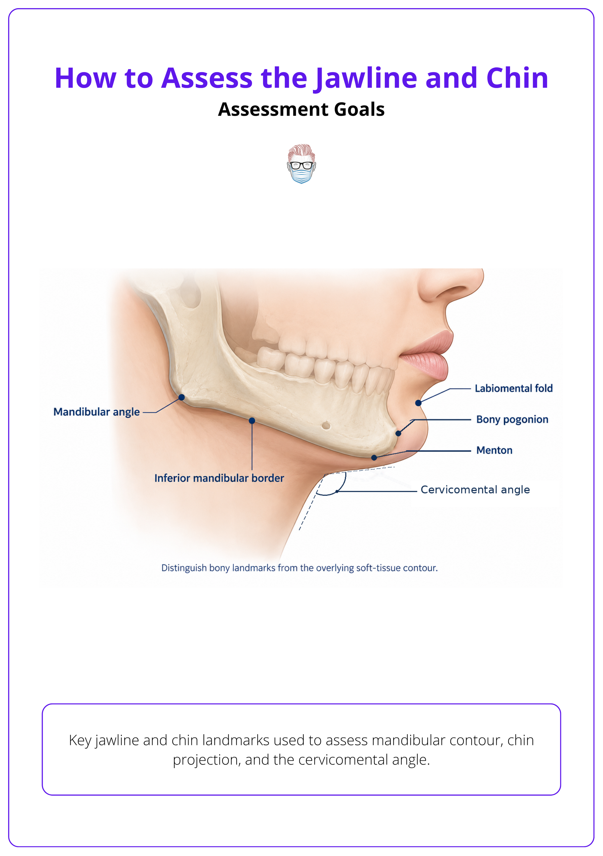
Assess the lower face as part of the whole face, not as an isolated contour problem. The aim is to identify what is anatomical, what is dental, what is ageing-related, and what treatment can realistically change.
Whole-Face Analysis
Start with global facial shape, balance, and symmetry before judging the jawline or chin alone.
- Facial balance: Assess the lower third in relation to the midface, nose, lips, and neck 1.
- Baseline traits: Documenting facial traits affects diagnosis, treatment planning, option selection, and outcome quality 2.
- Individual goals: Avoid applying one universal jawline ideal; match goals to facial structure and patient priorities 3.
Chin Position
Decide whether the chin supports or disrupts facial balance, especially when dentofacial deformity is possible.
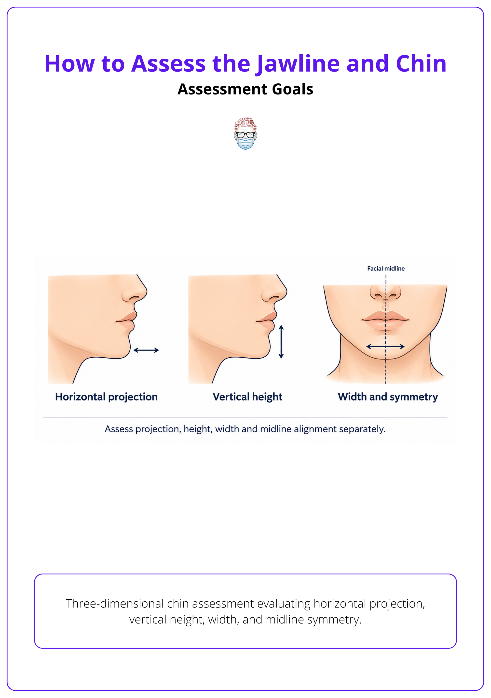
- Projection: Assess pogonion position relative to the lips, nose, and cervical contour.
- Vertical height: Separate a short chin from horizontal retrusion; they need different solutions.
- Width: Judge lower-face width against bizygomatic width, gonial width, and facial sex characteristics.
- Dentofacial clue: In dentofacial deformity, chin position may camouflage or exaggerate skeletal imbalance 4.
Treatment Direction
Define whether the problem is contour, volume, skeletal position, occlusion, or ageing before discussing treatment.
- Recordable features: Include chin projection, chin length and width, labiomental sulcus, mandibular angle, prejowl sulcus, jowls, and cervicomental angle 1.
- Grading: Use validated grading tools where possible so baseline severity and change can be compared consistently 5.
- Severity tracking: Chin retrusion and jawline sagging may be graded with 5-point photonumeric scales for objective baseline assessment 5.
- Suitability: Mild contour deficiency may suit camouflage; malocclusion or major skeletal discrepancy needs referral.
Applied Anatomy
Planning depends on the mandible, pogonion, dental relationship, labiomental fold, prejowl sulcus, soft-tissue envelope, facial vessels, mental nerve, marginal mandibular nerve, and ageing changes.
The lower face is a combined skeletal, dental, muscular, neurovascular, and skin-envelope assessment. Anatomy matters because the same visible deformity may need filler, implant, osteotomy, orthognathic correction, or neck treatment.
Skeletal Landmarks
Identify the fixed bony framework before assessing soft-tissue contour.
- Pogonion: The most anterior chin point guides perceived projection.
- Menton: The lowest midline point helps assess vertical chin height.
- Mandibular border: Palpate continuity from chin to angle; deficiency may be skeletal or soft-tissue masking.
- Assessment record: Separate projection, width, mandibular angle, prejowl sulcus, jowls, and cervicomental angle 1.
Soft-Tissue Envelope
Separate skeletal deficiency from soft-tissue descent, thickness, laxity, and muscular activity.
- Labiomental fold: A deep fold may accentuate retrusion; a shallow fold changes reduction planning.
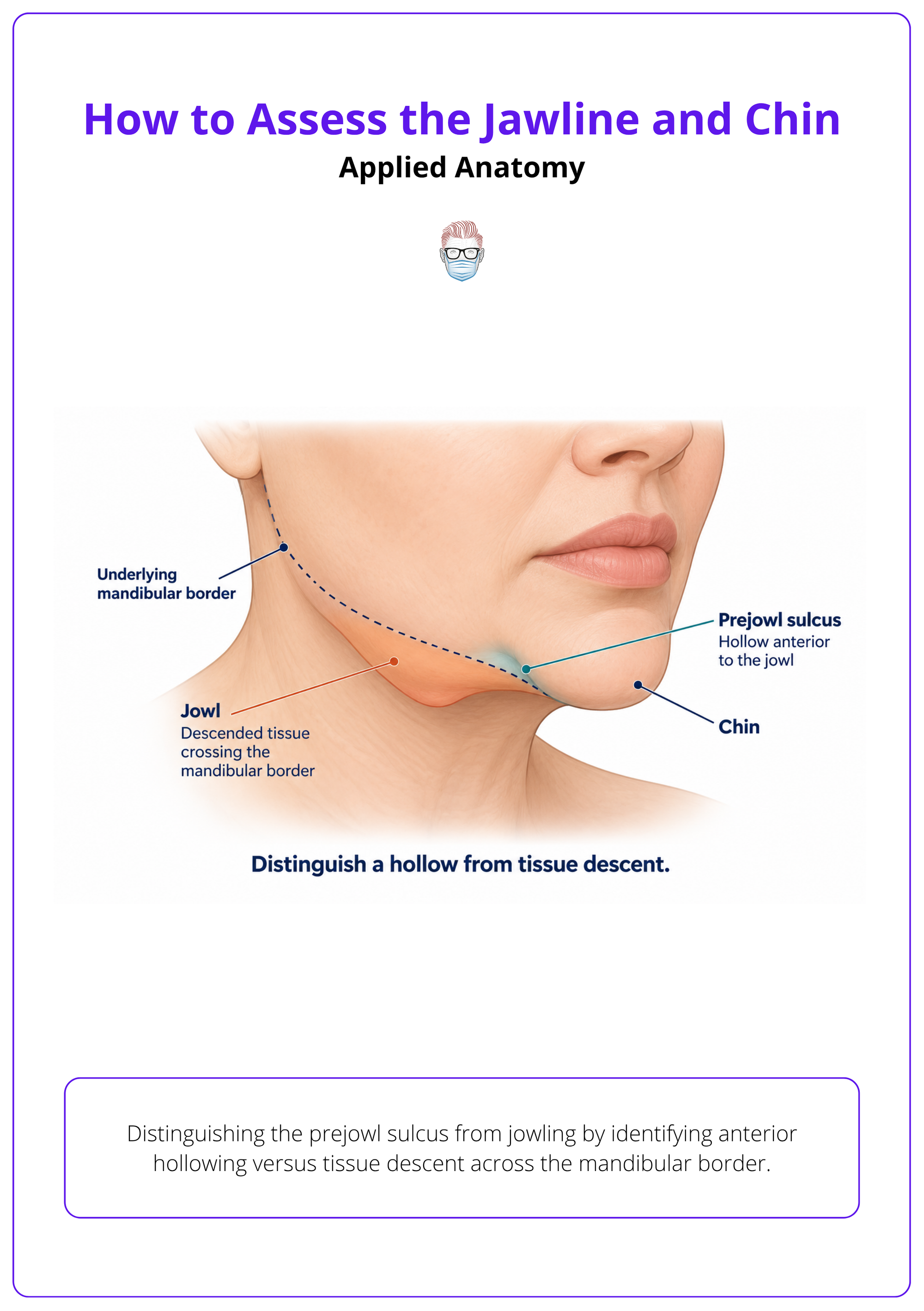
- Prejowl sulcus: Often reflects mandibular support loss, soft-tissue descent, or both.
- Platysma: Dynamic platysma contraction can reduce jawline definition and create vertical neck bands 6.
- Layer planning: Filler assessment should distinguish skeletal from soft-tissue landmarks to guide depth and contour 3.
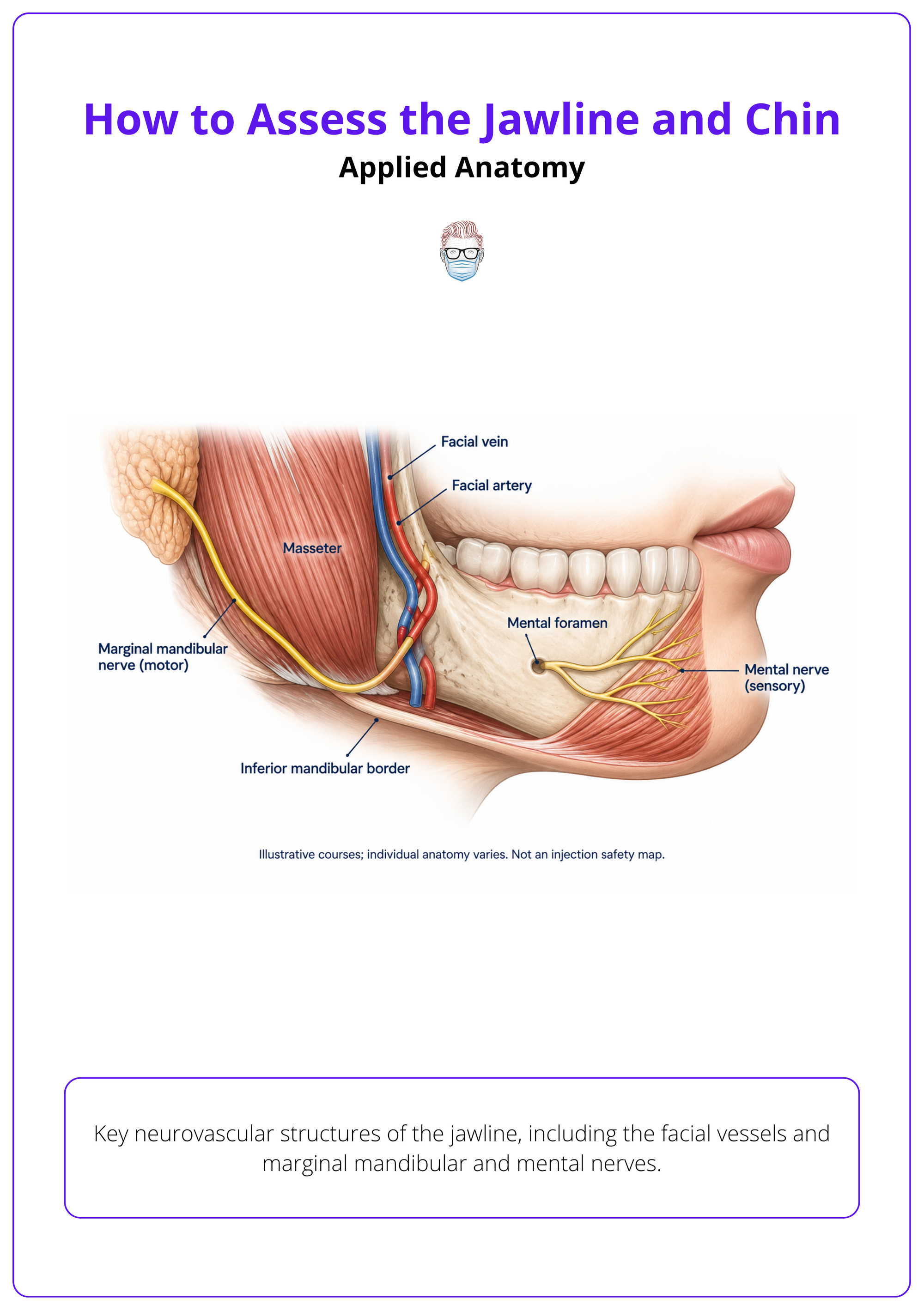
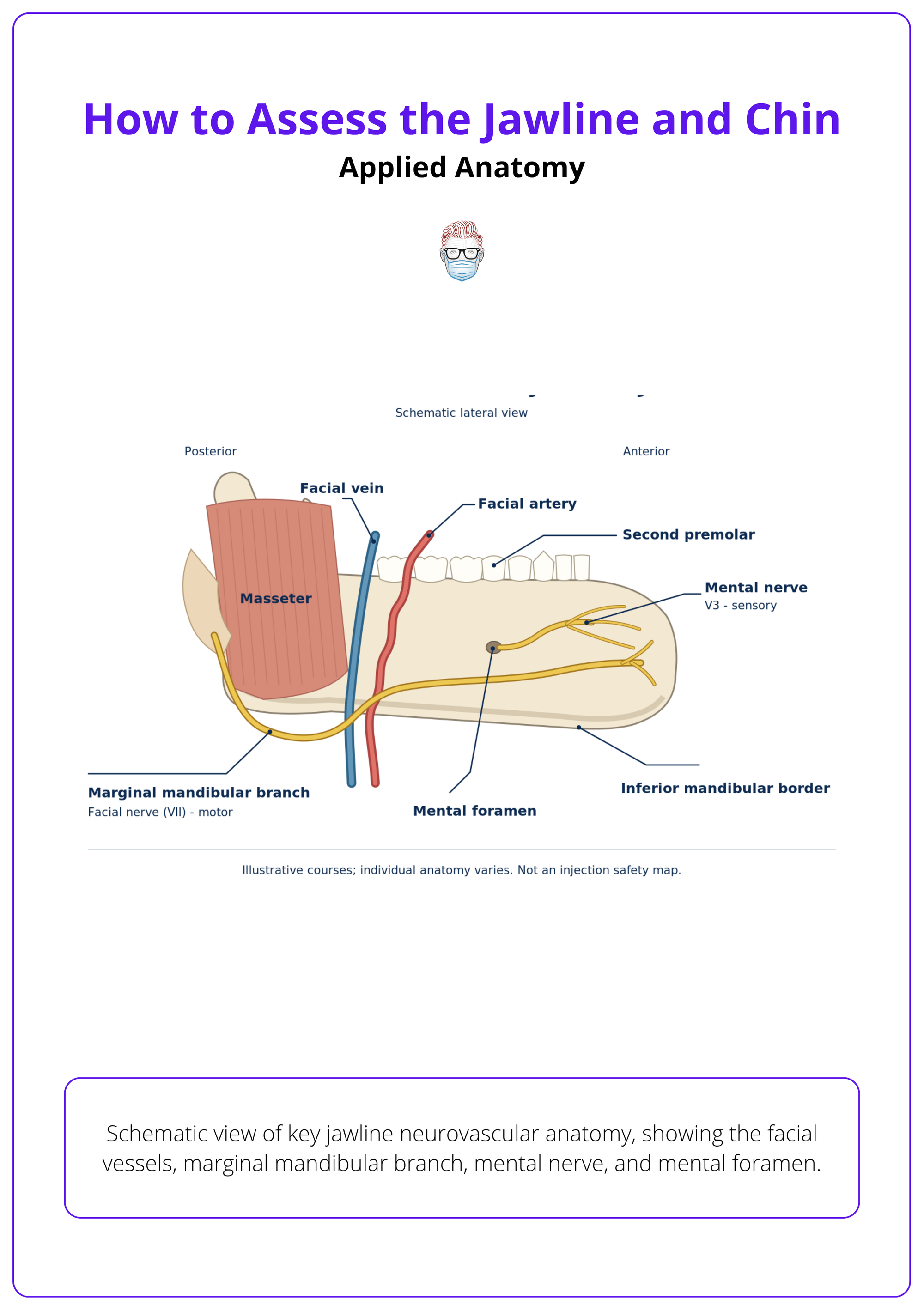
Danger Structures
Map nerves and vessels before injections, dissection, or osteotomy planning.
- Mental nerve: Localise the mental foramen region, classically around the second inferior premolar axis, because the mental nerve exits here and can be painful or vulnerable during chin and prejowl procedures 1.
- Mental vessels: The mental arteries emerge laterally near the second inferior premolar axis 1.
- Facial vessels: Lower-face filler planning requires awareness of facial artery, vein, and facial nerve location and depth 1.
- Marginal mandibular nerve: Consider it when assessing jowls, neck laxity, and surgical jawline access.
Operative Attachments
For osseous chin movement, soft-tissue attachment preservation protects segment support and vascularity.
- Anterior attachments: Avoid unnecessary stripping around planned genioplasty segments.
- Lingual attachments: Preserve deep attachments where possible during osteotomy planning 4.
- Lateral injection risk: Masseter-region and lateral jawline planning should account for variable transverse facial artery pathways 7.
Clinical Assessment
Assessment requires focused history, upright static and dynamic examination, chin projection and vertical analysis, asymmetry assessment, occlusion screen, palpation, standardised photography, and referral triggers.
Do not choose treatment from a lateral photograph alone. The clinical assessment should identify the dominant anatomical problem and expose dental, skeletal, dynamic, or soft-tissue factors that would make simple contour treatment unsafe or disappointing.
Focused History
History should identify aesthetic priorities, functional clues, prior treatment, and factors that alter sequencing.
- Presenting concern: Ask whether the patient sees retrusion, width, jowls, asymmetry, neck fullness, or poor definition.
- Dental history: Record orthodontics, extractions, restorations, jaw surgery, trauma, TMJ symptoms, and recent dental work.
- Treatment history: Ask about filler, toxin, implants, threads, liposuction, facelifting, and complications.
- Functional symptoms: Screen for changes in occlusion, chewing difficulty, speech issues, lip incompetence, snoring, and sleep-apnoea symptoms.
Static Examination
Examine upright at rest before asking for animation or planning treatment.
- Views: Assess frontal, profile, and oblique views in natural head position.
- Core measurements: Record chin projection, length, width, labiomental sulcus, mandibular angle, prejowl sulcus, jowls, and cervicomental angle 1.
- Structural planning: For possible genioplasty, assess symmetry, vertical dimension, width relative to face shape, and inferior mandibular border canting 8.
- Lip posture: Assess with lips relaxed so muscular compensation does not distort lip incompetence or soft-tissue prediction 4.
Dynamic Examination
Animation reveals muscular compensation, platysma activity, and lower-lip problems hidden at rest.
- Smile: Look for chin pad strain, lower-lip asymmetry, mentalis hyperactivity, and dental show.
- Pout and closure: Identify lip incompetence, mentalis dimpling, and effortful seal.
- Platysma activation: Ask for neck contraction to assess bands and jawline blunting.
- Upright documentation: Examine at rest and during animation, then document frontal, lateral, and oblique photographs 1.
Occlusal Screen
Aesthetic treatment should pause when the bite suggests skeletal or dental pathology.
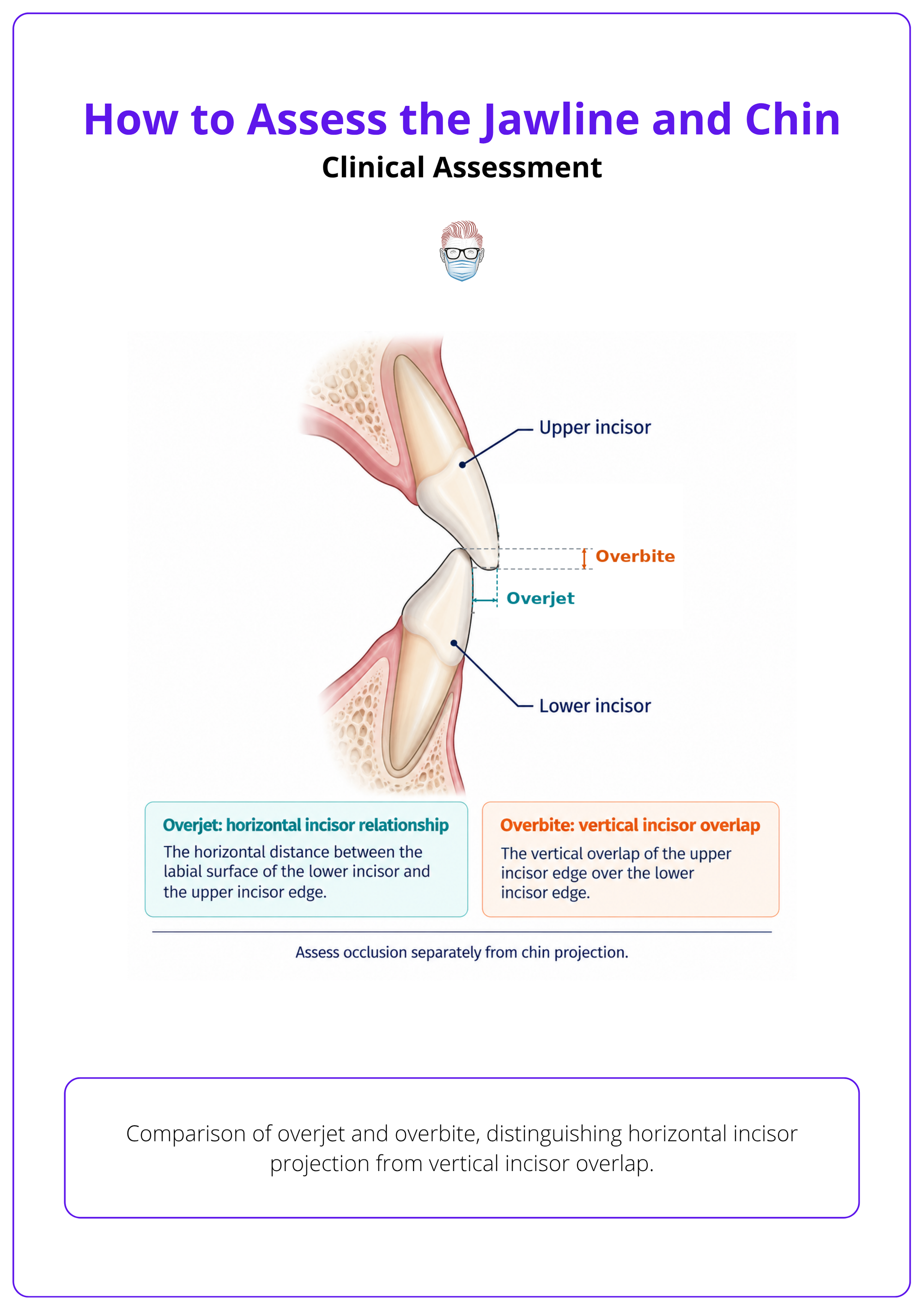
- Occlusion: Screen molar and canine relationship, overjet, overbite, crossbite, open bite, and midline shift.
- Dental interference: Palpation and dental assessment matter because orthognathic or odontological problems may need management first 1.
- Referral triggers: Refer for Class II or III discrepancy, chin compensation, facial asymmetry with occlusal cant, or unexplained bite change.
- Cleft history: Prior cleft repair or maxillary hypoplasia changes interpretation and usually needs specialist input.
Photography
Photographs are part of assessment, consent, planning, and medicolegal documentation.
- Standard set: Take frontal, lateral, three-quarter, basal, or submental, and dynamic views where relevant.
- Plane control: Standardise frontal, lateral, and three-quarter views with the Frankfort horizontal plane 4.
- Medicolegal value: Standardised photographs support planning, documentation, and record keeping 4.
- Validated scale: In relevant patients, the Asian Chin Projection Scale can grade projection from 0 to 4 9.
Analysis and Classification
Organise findings into horizontal, vertical, transverse, soft-tissue, ageing, mandibular-angle, and dentofacial patterns because each pattern points to a different treatment pathway.
Classification prevents the common error of treating every lower-face concern as “needs jawline filler.” The useful question is not whether the chin is small, but in which vector, by what mechanism, and with what occlusal relationship.
Vector Analysis
Classify the chin as horizontal, vertical, transverse, or combined before proposing correction 4.
- Horizontal deficiency: Microgenia or retrusion reduces profile support and may deepen the labiomental fold.
- Horizontal excess: Macrogenia or protrusion may dominate the lower third and sharpen the labiomental fold.
- Vertical deficiency: A short chin reduces lower-third height and may create a compressed mouth-chin unit.
- Vertical excess: A long chin may need reduction planning rather than augmentation.
Pattern Classification
Link the visible deformity pattern to the likely treatment implication before choosing a modality 4.
| Deformity pattern | Typical finding | Treatment implication |
|---|---|---|
| Microgenia / retrusion | Weak pogonion, poor profile support | Filler, implant, or advancement genioplasty |
| Vertical excess | Long lower third, elongated chin | Reduction genioplasty or orthognathic review |
| Transverse asymmetry | Chin point deviation, border cant | Osteotomy planning or orthognathic referral |
| Prejowl sulcus | Hollow anterior to jowl | Camouflage, implant wing support, lift strategy |
| Mandibular angle deficiency | Narrow lower face, weak gonial contour | Filler, implant, or skeletal contouring assessment |
| Dentofacial discrepancy | Malocclusion, overjet, crossbite, cant | Orthodontic–orthognathic referral |
Soft-Tissue Patterns
Separate contour loss from tissue descent because adding volume to descended tissue can worsen heaviness.
- Prejowl hollow: Identify whether it is a true hollow, jowl overhang, or mandibular border deficiency.
- Jowling: Look for ptotic tissue crossing the mandibular border rather than lack of bone alone.
- Neck laxity: Cervicomental angle, submental fat, and platysma bands may dominate jawline definition.
- Skin quality: Thin or lax skin limits camouflage and makes overfilling more visible.
Dentofacial Patterns
Malocclusion changes the diagnosis from aesthetic contour concern to facial skeletal assessment.
- Class II pattern: Retrognathia or microgenia may coexist with lip incompetence and deep bite.
- Class III pattern: A prominent chin may reflect maxillary deficiency, mandibular excess, or both.
- Cleft-related deformity: Do not assume cleft-related Class III patterns match noncleft Class III anatomy because maxillary and mandibular deficiencies may be greater 10.
- Midline discrepancy: Chin deviation with dental midline shift suggests skeletal asymmetry rather than filler asymmetry.
Severity Grading
Use structured scales when severity tracking or treatment comparison matters.
- Photonumeric scales: Chin retrusion and jawline sagging can be graded with validated 5-point scales 5.
- Ordered severity: These frameworks grade findings from none to very severe 5.
- Clinical use: Validated scales help standardise assessment in practice or research 5.
- Practical limit: A scale supports judgement; it does not replace occlusal and skeletal analysis.
Investigations and Referral Thresholds
Photographs may be enough for mild isolated contour concerns, but malocclusion, skeletal discrepancy, asymmetry, surgical planning, restorative dental issues, or airway symptoms should trigger dental, orthodontic, cephalometric, CBCT, CT, or sleep referral.
Investigations should answer a management question. Do not image every jawline patient, but do not camouflage a skeletal or dental problem because imaging was inconvenient.
Photographic Assessment
Use photographs alone only when the concern is mild, isolated, and clinically consistent with camouflage treatment.
- Adequate cases: Mild chin contour, mild prejowl hollowing, or soft-tissue asymmetry may not need imaging.
- Minimum record: Clinical assessment should still record projection, length and width, labiomental sulcus, mandibular angle, prejowl sulcus, jowls, and cervicomental angle 1.
- Deferral: If the examination does not explain the deformity, do not proceed from photographs alone.
- Severe laxity: Severe laxity, contour loss, or volume deficiency should prompt surgical referral rather than injectable-only treatment 1.
Dental Imaging
Dental or orthodontic assessment is needed when the bite, teeth, or restorative plan may drive the lower-face appearance.
- Dental priority: Treat dental and orthognathic problems first when they may interfere with jawline or chin results 1.
- Cephalometry: Profile analysis for restorative or full-arch aesthetic planning may include Holdaway angle, nasolabial angle, labiomental sulcus, and lower lip–pogonion relationships 11.
- Panoramic radiograph: Consider when dental status, mandibular pathology, implants, or planned surgery affects treatment.
- Sleep screen: Refer for sleep evaluation when symptoms suggest obstructive sleep apnoea or airway planning is relevant.
Surgical Planning
Advanced imaging is justified when skeletal movement, asymmetry, or orthognathic planning will change the operation.
- Virtual planning: Complex or subtle chin-position problems may warrant virtual surgical planning rather than visual assessment alone 4.
- Orthognathic genioplasty: CAD/CAM planning may be considered to improve accuracy and safety in orthognathic-context genioplasty 8.
- CBCT: CBCT-based 3D landmark assessment can support jaw-relationship measurement in skeletal dentofacial or cleft-related discrepancies 10.
- Class III threshold: Skeletal Class III features should prompt orthodontic–orthognathic referral because at least one-third may require surgery for aesthetic and functional correction 12.
Treatment Suitability Framework
Match treatment to anatomy: filler or toxin for selected mild contour and dynamic problems, implant for isolated horizontal augmentation, sliding genioplasty for skeletal vector change, orthognathic referral for malocclusion, and neck contouring for laxity or submental fullness.
Treatment selection follows diagnosis. The safest answer is often not “more volume,” but the option that corrects the dominant structural, dental, dynamic, or soft-tissue problem with the least distortion.
Non-Operative Management
Use injectables only when the deformity is mild, soft-tissue compatible, and not primarily occlusal or skeletal.
- Filler suitability: Injectable contouring suits light-to-moderate contour loss, volume loss, or constitutional lower-face deficiency 1.
- Product choice: Use high-viscosity filler for deep support and softer product for surface refinement when filler is appropriate 3.
- CaHA/CMC: Calcium hydroxylapatite/carboxymethylcellulose is a supported option for jawline contouring in suitable patients 13.
- Chin caution: Chin and prejowl CaHA/CMC treatment may help, but evidence should be framed less definitively than for jawline indications 13.
Neuromodulator Use
Use toxin for selected platysma-driven jawline blunting, not for heavy jowls, fat, or skeletal deficiency.
- Selection: OnabotulinumtoxinA evidence applies to adults with BMI 18–30 kg/m2 and bilateral moderate-to-severe platysma prominence at maximum contraction 6.
- Exclusions: Be cautious extrapolating results to excessive laxity, submental fat, jowls, or patients at increased toxin risk 6.
- Assessment rule: If the jawline improves when platysma relaxes, toxin may be logical; if tissue hangs, lifting or reduction may be needed.
Implant Augmentation
Consider a chin implant when the problem is mainly isolated horizontal deficiency with acceptable occlusion and vertical height.
- Best fit: Mild-to-moderate microgenia, stable bite, and a need for projection without vertical movement.
- Limitations: Implants do not correct significant vertical excess, asymmetry, occlusal abnormality, or mandibular border cant.
- Structural choice: Suitability should distinguish implant augmentation from osteotomy-based repositioning 4.
- Anaesthesia planning: Alloplastic chin implantation may be feasible with local anaesthesia and sedation in appropriate patients 4.
Osseous Genioplasty
Choose sliding genioplasty when the chin needs skeletal movement in horizontal, vertical, or transverse vectors.
- Vector correction: Osteotomy designs can address horizontal, vertical, and transverse or asymmetric movement requirements 4.
- Preoperative measures: Assess symmetry, vertical dimension, width, and inferior mandibular border canting before selecting the movement 8.
- Anaesthesia: Osseous genioplasty requires anaesthesiology involvement and more formal perioperative planning 4.
- Medical suitability: ASA III–IV patients may be unsuitable for outpatient genioplasty and need higher-acuity planning 4.
Referral And Adjuncts
Refer when the chin is a manifestation of bite, jaw, airway, or neck pathology rather than an isolated contour problem.
- Orthognathic pathway: Malocclusion, facial asymmetry with cant, Class II or III discrepancy, or cleft history should trigger referral.
- Neck contouring: Submental fat, skin laxity, and platysma bands may need liposuction, neck lift, or platysma treatment.
- Surgical sequencing: Treat dental and skeletal drivers before camouflage if they determine facial balance.
- Injection safety: If filler remains appropriate, review facial artery and branch risk before selecting planes and volumes 3.
Pitfalls and Safety Checks
Poor outcomes usually follow missed malocclusion, overfilling skeletal deficiency, ignoring asymmetry or vertical problems, treating jowls as hollows, missing nerve or vascular danger zones, or inadequate documentation.
Most errors occur before treatment starts. The safety check is to ask whether the planned treatment corrects the cause of the finding or simply hides it temporarily.
Diagnostic Pitfalls
Do not treat a lower-face contour complaint until skeletal, dental, soft-tissue, and dynamic causes have been separated.
- Missed bite: Malocclusion or chin compensation can make filler or implants look wrong after later jaw correction.
- Vertical error: Horizontal augmentation does not fix a long chin or short lower third.
- Asymmetry: Midline deviation, canting, and mandibular border asymmetry need documentation before consent.
- Platysma: Ignoring contraction can miss a major cause of jawline blunting and vertical neck bands 6.
Injection Safety
Filler planning must prioritise vascular risk, tissue plane, and signs of ischaemia.
- Vascular review: Assess the facial artery and its branches before chin or jawline filler 3.
- Technique: If proceeding, inject slowly, with low pressure, in the intended plane 1.
- Ischaemia: Stop and manage urgently if pain or blanching suggests local ischaemia 1.
- Risk line: For lateral face or jawline injections, treat the tragus-to-outer-upper-lip line as a risk area 7.
Treatment Errors
Avoid volume placement or osteotomy planning that worsens the original contour problem.
- Jowl filling: Do not place filler directly into bulky ptotic jowl tissue when restoring the mandibular line 1.
- Dental timing: Postpone lower-face filler after dental procedures within the previous 15 days or active dental problems 1.
- Blindness mechanism: Lateral jawline injections carry a rare blindness-risk mechanism with type III transverse facial artery anatomy 7.
- History check: Document allergies, prior injectable treatments, medication use, and relevant medical history before injection 1.
Operative Safety
Surgical planning should protect roots, nerves, contour, and soft-tissue support.
- Osteotomy level: Intraoral genioplasty osteotomy should be at least 5 mm below incisor roots and the mental foramen 4.
- Labiomental fold: Reconsider horizontal reduction genioplasty in a minimal labiomental fold because contour flattening may result 4.
- Mental nerve: Localise the foramen and counsel about temporary or persistent sensory symptoms.
- Implant sizing: Oversized implants can masculinise, distort the fold, or expose pre-existing asymmetry.
Counselling and Documentation
Patients should understand proportional diagnosis, treatment limits, asymmetry, staged care, maintenance needs, specific risks, and the recorded baseline findings before any jawline or chin intervention.
Counselling should show that the assessment has identified the cause of the concern, not just the requested treatment. Documentation should protect the decision pathway as much as the consent form.
Patient Counselling
Explain what treatment can improve, what it cannot camouflage, and when staged care is safer.
- Proportional diagnosis: Chin disharmony is assessed against the lower facial third and whole-face proportions 4.
- Expectation setting: Document the patient’s specific lower-face concerns and realistic goals before proposing treatment 1.
- Toxin counselling: In selected platysma-prominence patients, 61.2% reported satisfaction or high satisfaction at Day 14 after onabotulinumtoxinA versus 11.8% with placebo 6.
- Adverse events: The same trial reported TEAEs of 16.3% with onabotulinumtoxinA versus 19.9% with placebo 6.
Documentation
Record the baseline anatomy, occlusion screen, photographs, risks discussed, and shared decision.
- Baseline record: Include frontal, profile, oblique, and dynamic photographs where relevant.
- Occlusion: Document overjet, overbite, crossbite, midline shift, lip competence, and referral advice.
- Filler consent: Include uncommon but described risks such as infection, nodules, vascular compromise, ischaemia, tissue necrosis, and foreign body granulomas 11518.
- Follow-up: For filler patients, aftercare and follow-up assessment are part of maintaining the result 3.
References
[1] Eduardo. Reshaping the Lower Face Using Injectable Fillers.. Indian journal of plastic surgery : official publication of the Association of Plastic Surgeons of India. 2020. doi:10.1055/s-0040-1716185
[2] Arnett et al.. Facial keys to orthodontic diagnosis and treatment planning. Part I.. American journal of orthodontics and dentofacial orthopedics : official publication of the American Association of Orthodontists, its constituent societies, and the American Board of Orthodontics. 1993. doi:10.1016/0889-5406(93)70010-L
[3] Hong et al.. Anatomic-Based Diagnosis and Filler Injection Techniques: Chin Augmentation and Jawline Contouring.. The Journal of craniofacial surgery. 2025. doi:10.1097/SCS.0000000000011075
[4] Degala et al.. Genioplasty - A Review\.. Nigerian journal of clinical practice. 2024. doi:10.4103/njcp.njcp\_24\_24
[5] Pooth et al.. Validated 5-point photonumeric scales for the assessment of the jowls and chin.. Journal of cosmetic dermatology. 2022. doi:10.1111/jocd.14661
[6] Shridharani et al.. Improving Neck and Jawline Aesthetics With OnabotulinumtoxinA by Minimizing Platysma Muscle Contraction Effects: Efficacy and Safety Results in a Phase 3 Randomized, Placebo-Controlled Study.. Aesthetic surgery journal. 2025. doi:10.1093/asj/sjae220
[7] Toure et al.. Transverse facial artery: Its role in blindness after cosmetic filler and botulinum toxin injections.. Journal of plastic, reconstructive & aesthetic surgery : JPRAS. 2021. doi:10.1016/j.bjps.2020.12.042
[8] Ramanathan et al.. Genioplasty in Contemporary Orthognathic Surgery.. Oral and maxillofacial surgery clinics of North America. 2023. doi:10.1016/j.coms.2022.06.009
[9] Suwanchinda et al.. A Validated Assessment Scale for Asian Chin Projection.. Dermatologic surgery : official publication for American Society for Dermatologic Surgery \[et al.]. 2019. doi:10.1097/DSS.0000000000001996
[10] Sinha et al.. Does cleft lip and palate affect the severity of malocclusion?. Clinical oral investigations. 2023. doi:10.1007/s00784-023-05345-z
[11] Orphanos. Facial Analysis: The Roadmap to Full-Arch Esthetic Success.. Oral and maxillofacial surgery clinics of North America. 2025. doi:10.1016/j.coms.2024.11.002
[12] Singh et al.. Individualized Surgical Orthodontic Rehabilitation of Class III Dentofacial Deformities.. International orthodontics. 2020. doi:10.1016/j.ortho.2019.08.022
[13] Guida et al.. A systematic review of Radiesse/calcium hydroxylapatite and carboxymethylcellulose: evidence and recommendations for treatment of the face.. International journal of dermatology. 2024. doi:10.1111/ijd.16888
