


Structure ageing face assessment from global impression to regional analysis, then separate volume deflation, tissue deterioration, and soft-tissue descent before discussing treatment .
Applied Facial Anatomy
Assessment should use a layered anatomical model of skin, subcutaneous fat, SMAS, deep fat, and periosteum or deep fascia because each layer changes treatment choice .
History and Suitability
Suitability assessment starts with the patient’s concerns and desired improvements, then checks medical risk, psychiatric history, medications, prior treatment, lifestyle, and consent capacity .
Systematic Clinical Examination
Examine and document the ageing face in standard conditions, at rest and animation, recording skin quality, volume loss, descent, asymmetry, proportions, and region-specific findings.
Diagnosis and Problem Prioritisation
Translate findings into dominant diagnoses such as skin change, volume loss, ptosis, skeletal deficiency, dynamic lines, laxity, or asymmetry, then rank them against the patient’s stated concern.
Investigations and Adjunctive Assessment
Use adjuncts selectively when they improve documentation, functional assessment, diagnosis, safety, or follow-up, not as substitutes for clinical judgement.
Treatment Planning Principles
Treatment planning should match the dominant ageing diagnosis to skin, volume, neuromodulator, energy-based, surgical, or combined strategies while preserving facial balance.
Documentation, Counselling, and Red Flags
Document baseline findings, photographs, patient goals, consent-relevant risks, psychological suitability, and red flags that require deferral, referral, safeguarding, or medical workup.
Ageing Face Assessment Framework
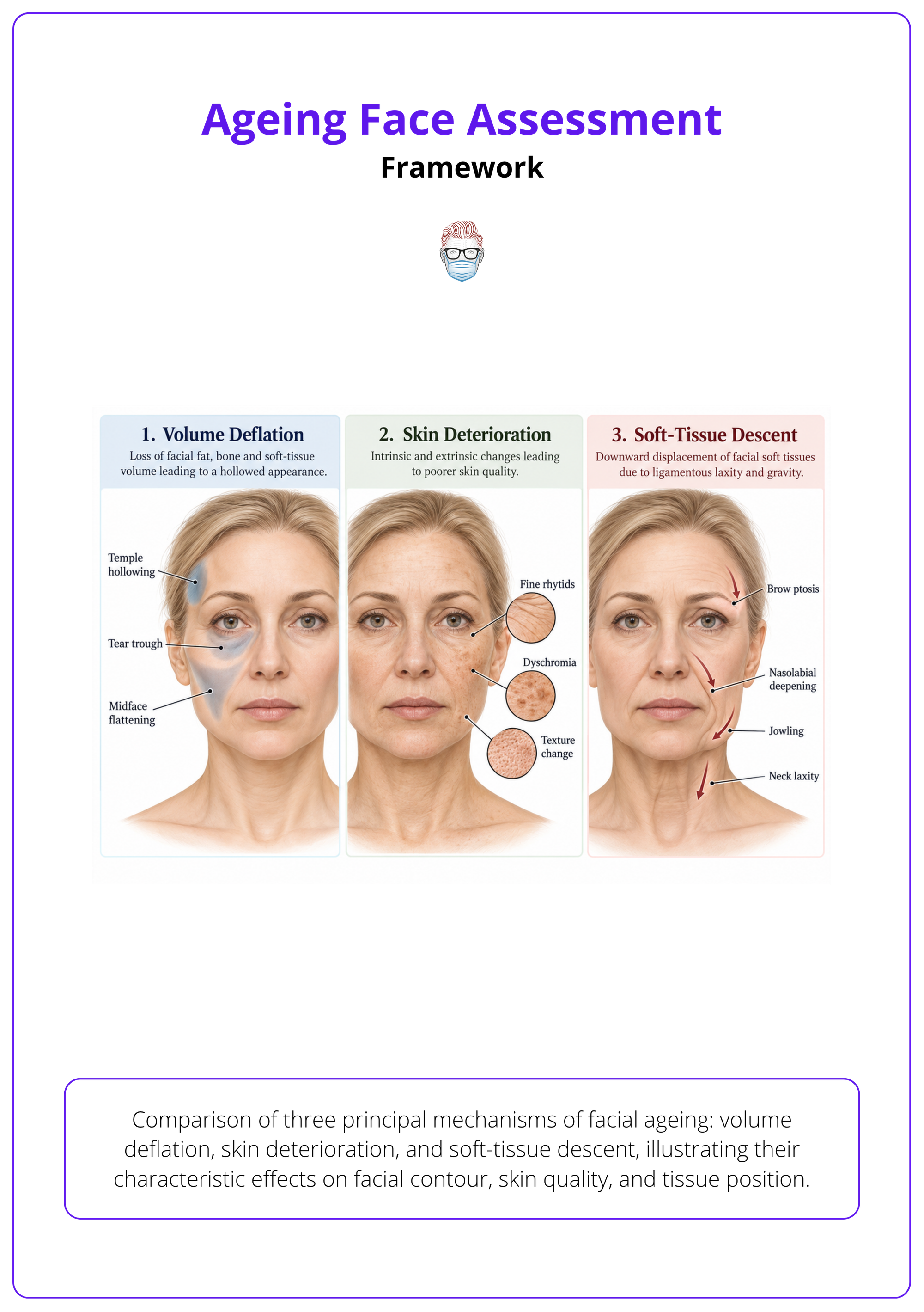
Structure ageing face assessment from global impression to regional analysis, then separate volume deflation, tissue deterioration, and soft-tissue descent before discussing treatment 1.
Ageing face assessment is an aesthetic diagnosis, not a menu of procedures. The aim is to identify what has changed, why it has changed, and which findings matter to the patient.
Aesthetic Diagnosis
Name the ageing mechanism before naming the treatment.
- Volume deflation: look for hollowing, contour loss, tear troughs, temple hollowing, and midface flattening.
- Tissue deterioration: assess skin quality, photodamage, rhytids, texture, laxity, and pigmentation.
- Soft-tissue descent: identify brow ptosis, malar descent, jowling, nasolabial deepening, and neck laxity.
- Dynamic contribution: separate lines present at rest from lines produced by mimetic animation.
Whole-Face Analysis
Assess the whole three-dimensional face before deciding which regions deserve intervention.
- Global impression: observe age, facial balance, expression, tiredness, heaviness, and symmetry.
- Regional survey: include upper, mid, lower, periocular, and perioral regions before prioritising treatment 2.
- Periorbital unit: assess brow, eyelid, temporo-orbital, and malar ageing as one unit, not isolated eyelid skin 3.
- Perioral unit: include the mouth and upper lip because perioral ageing contributes meaningfully to perceived age 4.
Skin Quality
Do not reduce facial ageing to wrinkle severity alone.
- Texture: record roughness, fine lines, pores, crepiness, and actinic change.
- Colour: assess dyschromia, erythema, sallowness, lentigines, and uneven tone.
- Hydration: include dryness as a visible skin-quality domain in ageing assessment 5.
- Elasticity: judge recoil and laxity because these influence non-surgical versus surgical planning.
Assessment Sequence
Use a numbered 1-5 sequence every time, so important findings are not missed.
| Step | Clinical question | Output |
|---|---|---|
| Global view | What is the dominant ageing pattern? | First impression and asymmetry |
| Patient concern | What does the patient want changed? | Priority list in the patient’s words |
| Regional analysis | Which facial units are affected? | Upper, mid, lower, perioral, neck findings |
| Mechanism | What explains the finding? | Skin, volume, descent, muscle, skeleton |
| Planning | What strategy fits the diagnosis? | Non-surgical, surgical, resurfacing, combined, or defer |
Patient Variation
Counsel against a single idealised ageing pattern.
- Sex differences: male and female facial skeleton, soft tissue, hairline, and brow position age differently.
- Ethnic variation: preserve identity-defining features unless the patient specifically asks otherwise.
- Ageing rate: ageing severity varies by individual, sex, and race or ethnicity, which should inform counselling 1.
- Treatment threshold: visible ageing alone is not an indication unless it matches the patient’s goals and safe options.
Applied Facial Anatomy
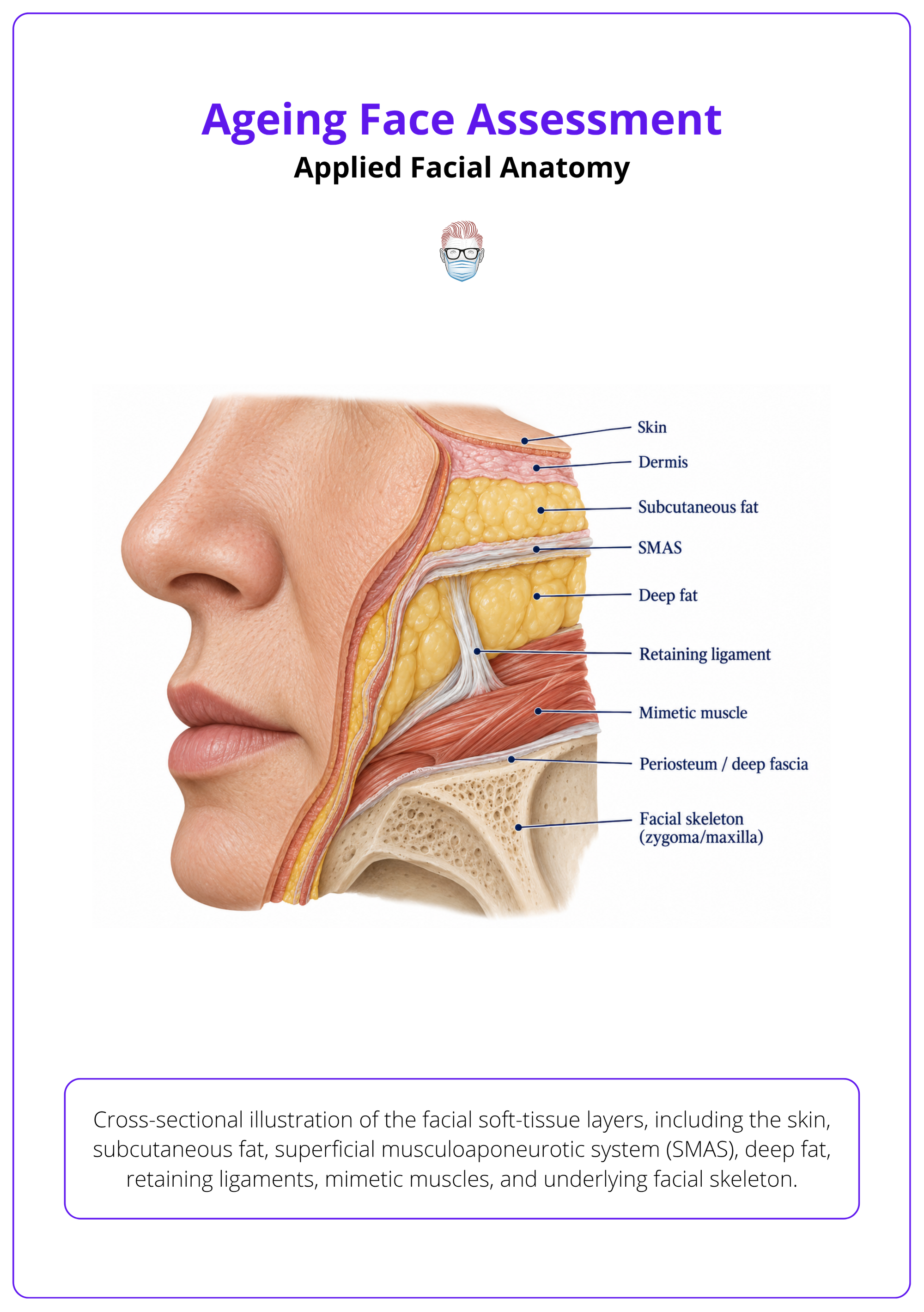
Assessment should use a layered anatomical model of skin, subcutaneous fat, SMAS, deep fat, and periosteum or deep fascia because each layer changes treatment choice 6.
Applied anatomy explains why one face needs skin treatment, another needs volume, and another needs lifting or skeletal support. Assessment should therefore identify the layer causing the visible problem.
Facial Layers
Link the visible deformity to the anatomical layer most responsible for it.
- Skin: photodamage, dyschromia, fine rhytids, texture change, and dermal thinning.
- Subcutaneous fat: superficial fullness, compartment descent, contour irregularity, and jowling.
- SMAS layer: soft-tissue support, facial lifting vectors, and surgical dissection relevance.
- Deep support: deep fat, periosteum, maxilla, mandible, and dental support affect facial projection.
Fat Compartments
Localise superficial and deep fat compartments before planning fillers, lifting, or contour correction.
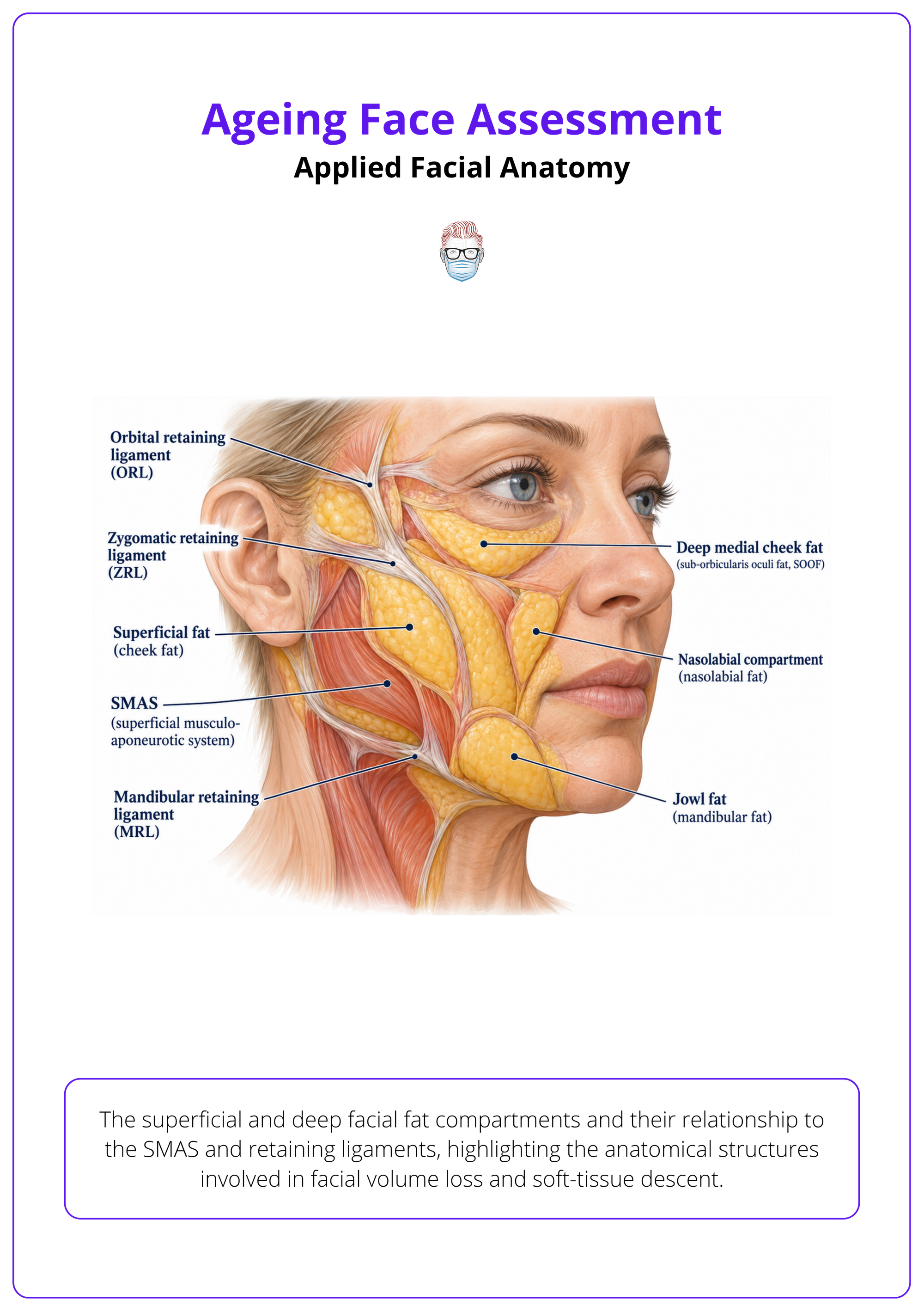
- Superficial fat: descent and compartment bulging contribute to nasolabial folds, jowls, and heaviness.
- Deep fat: deflation contributes to tear trough, midface flattening, and loss of anterior projection.
- Boundaries: compartment anatomy should be localised before minimally invasive rejuvenation to improve safety and effect 6.
- Regional interpretation: fullness may represent descent, true volume excess, oedema, or skeletal deficiency.
Retaining Ligaments
Use retaining ligaments as both ageing landmarks and operative risk landmarks.
- Assessment clue: ligamentous fixation creates folds, grooves, and transition points as mobile tissues descend.
- Planning relevance: operative planning should define the dissection plane and ligament release strategy 7.
- Nerve safety: retaining ligaments can guide safer dissection around facial nerve risk zones 7.
- Treatment limitation: fillers alone cannot reliably correct deformity driven by fixed descent and tethering.
Skeletal Support
Consider dental and skeletal support when soft-tissue ageing looks disproportionate.
- Midface support: maxillary retrusion or dental collapse can exaggerate nasolabial and perioral ageing.
- Mandibular support: chin and mandibular deficiency worsen jawline blunting and neck imbalance.
- Integrated assessment: dental, skeletal, and soft-tissue findings should be integrated when structural support affects facial shape 8.
- Documentation: record skin quality, laxity, photodamage, descent, and skeletal support because each may change procedural choice 9.
History and Suitability
Suitability assessment starts with the patient’s concerns and desired improvements, then checks medical risk, psychiatric history, medications, prior treatment, lifestyle, and consent capacity 9.
History decides whether to proceed, modify treatment, defer, or decline. A technically possible intervention is not automatically appropriate.
Aesthetic History
Clarify the patient’s concern before interpreting the face for them.
- Primary concern: ask what feature bothers them most and what change they would accept.
- Motivation: distinguish self-directed goals from external pressure, relationship conflict, or occupational anxiety.
- Prior treatment: document recent retinoids, fillers, radiofrequency, ultrasound, toxin, laser, peels, and dermabrasion because they alter findings and suitability 5.
- Identity: ask which facial or ethnic characteristics the patient wants preserved before planning aesthetic change.
Medical Risk
Identify conditions and medications that increase procedural risk or distort assessment.
- General health: screen for infection, inflammation, unstable disease, poor wound healing, and uncontrolled systemic illness.
- Medication risk: check anticoagulants, antiplatelets, NSAIDs, immunosuppression, isotretinoin exposure, and hypersensitivity history 5.
- Polypharmacy: medication history should identify polypharmacy and inappropriate prescribing because these are consistently associated with adverse outcomes in older people 10.
- Frailty trigger: age over 80 with subjective fatigue should prompt frailty-focused suitability screening before elective planning 11.
Psychological Suitability
Screen actively when the request, distress, or expectations seem disproportionate.
- Expectation mismatch: defer if the patient wants perfection, transformation, or guaranteed emotional change.
- BDD screening: patients seeking ageing-face treatment should be assessed face-to-face and screened for body dysmorphic disorder using approved scales when indicated 12.
- Psychiatric history: ask about depression, anxiety, eating disorder, self-harm, previous cosmetic dissatisfaction, and repeated consultations.
- Consent capacity: confirm the patient can understand options, uncertainty, risk, alternatives, and the possibility of no treatment.
Systematic Clinical Examination
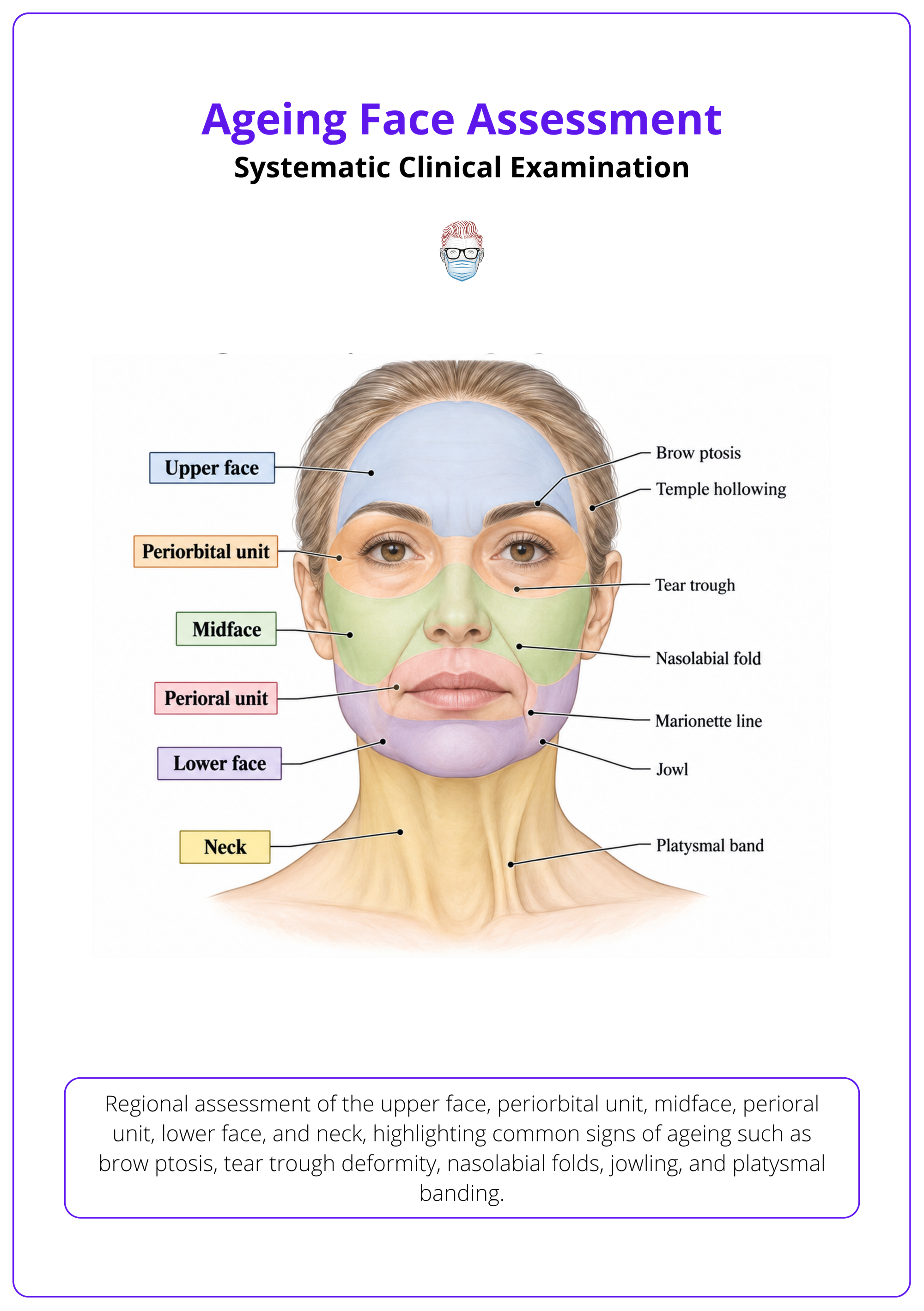
Examine and document the ageing face in standard conditions, at rest and animation, recording skin quality, volume loss, descent, asymmetry, proportions, and region-specific findings.
Examination should convert subjective ageing into observable findings. Use the same order each time: face first, regions second, measurements where they change decisions.
Examination Conditions
Standardise the setting before judging asymmetry, laxity, and surface quality.
- Position: examine seated upright with the head neutral and hair cleared from the forehead and ears.
- Lighting: use even frontal and oblique lighting; harsh shadows can overstate grooves and hollows.
- Repose: assess relaxed brow, eyelid aperture, oral commissures, jawline, and neck contour.
- Animation: examine frontalis, frown, smile, orbicularis, depressor anguli oris, mentalis, and platysma activity.
Facial Proportions
Record symmetry and proportions before focusing on isolated defects.
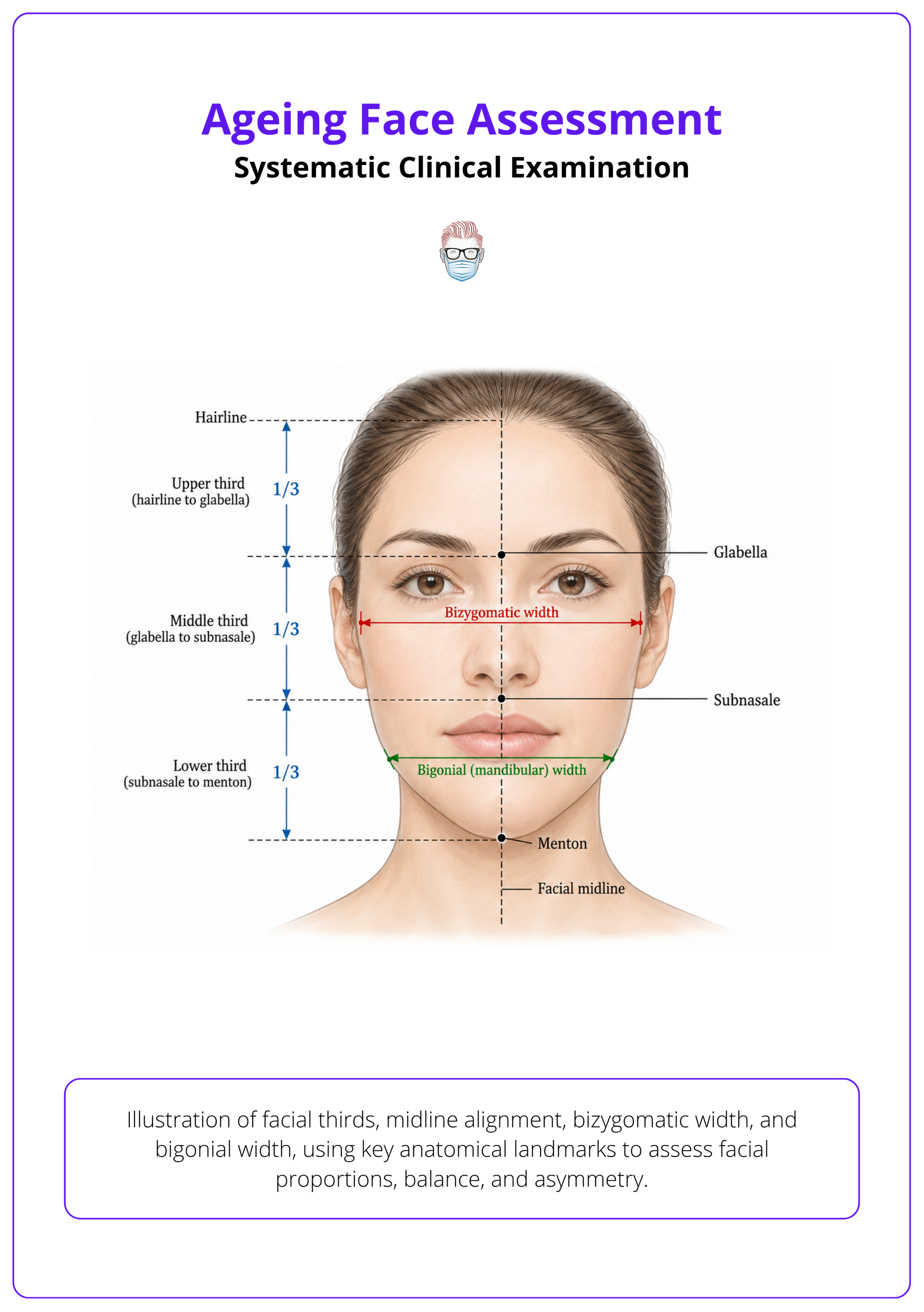
- Global measures: document symmetry, proportions, facial angles, and indices for the overall face 13.
- Vertical thirds: assess hairline to glabella, glabella to subnasale, and subnasale to menton balance.
- Transverse balance: compare bizygomatic width, mandibular width, chin point, and dental midline.
- Anthropometry: objective measurements support baseline analysis and comparison after treatment 13.
Skin Examination
Separate skin ageing domains because they respond to different treatments.
- Lines: record fine lines, static rhytids, deep folds, and dynamic wrinkles separately.
- Pigmentation: examine lentigines, melasma, post-inflammatory change, erythema, and uneven tone.
- Texture: document roughness, crepiness, thickness, pores, scars, and sebaceous quality.
- Photoageing: fine lines, wrinkles, uneven pigmentation, and roughness should be recorded as distinct findings 14.
Regional Examination
Examine each facial unit, then check whether neighbouring units explain the complaint.
- Upper face: brow height, brow shape, forehead lines, glabellar strain, temple hollowing, and hairline.
- Periorbital: document eyebrow ptosis, lateral temporo-orbital sagging, lid hooding, malar descent, crow’s feet, lid-cheek depth, and festoons 3.
- Perioral: assess philtral height, vermilion show, dental show, commissures, smoker’s lines, and lip ratios 4.
- Lower face and neck: record jowls, marionette lines, submental fullness, platysmal bands, and cervicomental angle.
Functional Measures
Add measurements when the finding may be functional, procedural, or follow-up relevant.
- Eyelid metrics: Margin Reflex Distance 1 (MRD1) can be recorded at baseline and follow-up when ptosis severity matters 15.
- Laxity metrics: brow position, submental area, and marionette-line displacement can quantify laxity for candidacy or response 16.
- Frailty screen: usual-pace gait speed is a quick, inexpensive examination measure in older aesthetic patients 11.
- Frailty domains: fatigue, stair climbing, ambulation, illness burden, and weight loss over 5% can be assessed during examination 11.
Diagnosis and Problem Prioritisation
Translate findings into dominant diagnoses such as skin change, volume loss, ptosis, skeletal deficiency, dynamic lines, laxity, or asymmetry, then rank them against the patient’s stated concern.
Problem prioritisation prevents treating the most visible feature while missing the dominant cause. The useful diagnosis is the one that changes planning.
Diagnostic Categories
Describe the ageing problem by mechanism rather than by the procedure that might treat it.
- Skin change: pigmentation, roughness, wrinkles, actinic damage, scars, and poor elasticity.
- Volume loss: temple hollowing, tear trough, midface flattening, perioral deflation, and mandibular contour loss.
- Ptosis: brow descent, malar descent, jowls, neck laxity, and platysmal banding.
- Skeletal deficiency: maxillary, mandibular, chin, dental, or occlusal support problems.
Regional Priorities
Organise diagnoses by facial region, then decide which region drives the complaint.
- Framework: prioritisation can be organised by forehead, periorbital area, midface, perioral area, and jawline 17.
- Midface: when present, diagnose deep-volume deflation with secondary superficial fat descent as a midface ageing driver 1.
- Lower face: prioritise jowl prominence and cervicomental angle increase when jawline definition is the main issue 1.
- Perioral and nasal: mouth lengthening, narrowing, nasal lengthening, widening, and tip descent may be age-related contributors 18 18.
Treatment-Relevant Diagnosis
The diagnosis should point to a strategy.
| Dominant finding | Likely diagnosis | Planning consequence |
|---|---|---|
| Fine lines and dyschromia | Skin-quality ageing | Skin care, resurfacing, energy, or peel discussion |
| Hollow temples or midface | Volume deflation | Volume restoration or structural support discussion |
| Heavy brow or jowls | Soft-tissue descent | Lifting strategy rather than filler alone |
| Lines only on animation | Dynamic rhytids | Neuromodulator discussion |
| Severe static folds | Combined skin, volume, and descent | Combined or staged plan |
| Disproportionate asymmetry | Structural, dental, neurological, or prior-treatment issue | Investigate or defer before treatment |
Functional Priorities
Do not miss functional pathology hidden inside aesthetic ageing.
- Upper eyelid ptosis: MRD1 of 0–2 mm was used to define moderate to severe ptosis in one cohort, supporting ptosis as a distinct diagnosis 15.
- Severe laxity: when severe skin laxity is dominant, surgical options should be prioritised over microfocused ultrasound 16.
- Platysma: platysma contraction should be diagnosed separately as a lower-face and neck ageing problem.
- Periorbital skin: wrinkles, hyperpigmentation, and photoageing should be separated because they are different treatable problems 20.
Patient Priorities
Rank objective findings against what the patient actually wants changed.
- Dominant concern: ask the patient to rank concerns before offering your own hierarchy.
- Mismatch: explain when the visible problem is not the anatomical cause.
- Skin priority: pigmentation, roughness, and wrinkles should be prioritised separately because different topical options fit different profiles 14.
- Volume priority: identify where volume restoration or contour enhancement is expected to give the greatest rejuvenative effect 2.
Investigations and Adjunctive Assessment
Use adjuncts selectively when they improve documentation, functional assessment, diagnosis, safety, or follow-up, not as substitutes for clinical judgement.
Most ageing face assessments need good history, examination, and photographs rather than extensive tests. Investigations are useful when they change the plan.
Photographic Assessment
Standardised images are the main adjunct for baseline comparison and treatment review.
- Clinical photographs: take frontal, oblique, lateral, smiling, and animation views when relevant.
- Serial review: baseline and serial two-dimensional photographs can support objective follow-up after microfocused ultrasound 16.
- Lip analysis: photographic ratios, caliper measurements, or three-dimensional imaging can quantify upper-lip ageing and treatment effect 4.
- Three-dimensional imaging: standardise capture with the patient seated, neutral-faced, and frontal when using 3D facial imaging 18.
Selected Testing
Order tests only when the finding may be functional, systemic, psychiatric, or technically complex.
- Visual fields: Goldmann perimetry can help when upper-eyelid morphology may affect the superior visual field 21.
- Orbital imaging: CT may be useful when thyroid eye disease is part of the eyelid-retraction differential 22.
- BDD tools: BDDQ-DV, BDDQ-AS, COPS, and BDSS can be considered when psychiatric suitability is uncertain 12.
- AI adjuncts: use AI cautiously in patients over 60 or underrepresented groups because training data limitations may reduce reliability 23 23.
Treatment Planning Principles
Treatment planning should match the dominant ageing diagnosis to skin, volume, neuromodulator, energy-based, surgical, or combined strategies while preserving facial balance.
Planning starts after diagnosis. Avoid moving directly from “looks tired” to blepharoplasty, filler, toxin, or facelift without identifying the anatomical cause.
Treatment Matching
Choose the strategy that treats the dominant anatomical problem.
- Assessment inputs: integrate facial volumes, lines, skin quality, validated scales, and standardised photography when selecting and evaluating treatment 17.
- Expectation setting: align goals with ageing severity, skin-envelope elasticity, and volume loss before offering treatment 2.
- Balance: prioritise harmonious facial balance rather than correcting isolated signs without context 1.
- Individualisation: combine subjective goals with objective anthropometric analysis to personalise surgical or non-surgical planning 13.
Non-Operative Management
Use non-surgical treatment when the problem is mild, reversible, dynamic, skin-quality driven, or volume-related.
- Core options: injectable soft-tissue fillers and neurotoxins are common non-surgical facial rejuvenation options 24.
- Skin laxity: microfocused ultrasound is best planned for mild-to-moderate facial skin laxity rather than advanced laxity 16.
- Modest goals: radiofrequency should be framed as a modest rejuvenation option for patients seeking modest improvement 25.
- Biostimulation: poly-L-lactic acid (PLLA) effects may last up to 25 months and CaHA effects 12 to 18 months when counselling injectable biostimulation 26.
Surgical And Combined Strategy
Escalate when the diagnosis is structural, severe, redundant, functional, or unlikely to respond safely to non-surgical treatment.
- Skin excess: redundant eyelid skin, severe neck laxity, jowling, and marked tissue descent often need surgical discussion.
- Upper lip: balance longer-lasting surgical lip lift and inevitable scarring against temporary minimally invasive options 4.
- Skeletal support: orthodontic or orthognathic intervention may be needed before or alongside cosmetic procedures when support is deficient 8.
- Combination planning: combine modalities only when each treats a separate diagnosis, such as skin quality, volume, and descent.
Documentation, Counselling, and Red Flags
Document baseline findings, photographs, patient goals, consent-relevant risks, psychological suitability, and red flags that require deferral, referral, safeguarding, or medical workup.
Good documentation protects the patient and the clinician. It should show what was assessed, what was diagnosed, what was discussed, and why the plan is appropriate or deferred.
Clinical Record
Record enough detail for another clinician to understand the baseline face and the decision.
- Photographs: standardised photographic documentation should form part of the record and support outcome assessment 9.
- Patient goals: document the patient’s own words, priority concerns, and unacceptable outcomes.
- Risk review: counselling should explicitly review medical and psychiatric history, medication use, and personal expectations 27.
- Objective grading: photographic wrinkle grading can support documentation because it correlated with profilometry-measured wrinkle depth in one study 28.
Patient Counselling
Explain what the finding means, what treatment can and cannot do, and what trade-off follows.
- Expectation control: avoid promising youthfulness, symmetry, emotional change, or permanent correction.
- Botulinum toxin: upper-face toxin counselling can include that most reported complications are mild and transient 29.
- Microfocused ultrasound: counselling should mention uncommon adverse effects occurred in 2% of treated patients in one series 16.
- Lip lift: discuss visible incision scarring because it was the most common long-term complication in one review, reported at 4.36% 4.
Red Flags
Defer or escalate when the assessment suggests psychological, medical, safeguarding, or diagnostic risk.
- BDD concern: repeated consultations for the same concern or psychiatric comorbidities should be treated as red flags 27.
- Screening tools: validated cosmetic-setting BDD tools can support referral decisions when concern persists 12.
- Safeguarding: do not ignore elder abuse concerns; pooled prevalence was 27.6% across 94 studies 30.
- Thyroid eye disease: document disease phase and thyroid status before drawing conclusions from eyelid measurements.
Ethical Documentation
Preserve identity, autonomy, and diagnostic uncertainty in the record.
- Cultural features: record features the patient wants preserved as well as those they want changed.
- Simulation limits: if outcome simulation is used, document that biased systems may alter rendered ethnicity 23.
- AI safeguards: when AI assists planning, document safeguards against undervaluing ethnic features 23.
- No-treatment decision: document when treatment is declined, deferred, or redirected because risk outweighs benefit.
References
[1] Swift et al.. The Facial Aging Process From the "Inside Out".. Aesthetic surgery journal. 2021. doi:10.1093/asj/sjaa339
[2] Murad et al.. Appropriate Selection and Application of Nonsurgical Facial Rejuvenation Agents and Procedures: Panel Consensus Recommendations. Aesthetic Surgery Journal. 2010. doi:10.1093/asj/30.1\_Supplement.36S
[3] Pascali et al.. The Temporal Subcutaneous Brow Lift with Orbicularis Oculi Muscle Elastic Flap: Technical Considerations, Systematic Review, and Terminology Standardization.. Facial plastic surgery : FPS. 2023. doi:10.1055/a-1953-2304
[4] Zhao et al.. Different Techniques and Quantitative Measurements in Upper lip lift: A Systematic Review\.. Aesthetic plastic surgery. 2023. doi:10.1007/s00266-023-03302-5
[5] Rattanawiwatpong et al.. Anti-aging and brightening effects of a topical treatment containing vitamin C, vitamin E, and raspberry leaf cell culture extract: A split-face, randomized controlled trial.. Journal of cosmetic dermatology. 2020. doi:10.1111/jocd.13305
[6] Cotofana et al.. Anatomy of the Facial Fat Compartments and their Relevance in Aesthetic Surgery.. Journal der Deutschen Dermatologischen Gesellschaft = Journal of the German Society of Dermatology : JDDG. 2019. doi:10.1111/ddg.13737
[7] Alghoul et al.. Retaining ligaments of the face: review of anatomy and clinical applications.. Aesthetic surgery journal. 2013. doi:10.1177/1090820X13495405
[8] Catherine et al.. Rejuvenation of the Ageing Face and the Role of Orthodontics: Guidelines for Management. Journal of Orthodontics. 2022. doi:10.1177/14653125221093390
[9] James et al.. Preoperative Assessment of the Aging Patient. Clinics in Plastic Surgery. 2006. doi:10.1016/j.cps.2006.02.004
[10] Davies et al.. Adverse Outcomes of Polypharmacy in Older People: Systematic Review of Reviews.. Journal of the American Medical Directors Association. 2020. doi:10.1016/j.jamda.2019.10.022
[11] Abellan et al.. The I.A.N.A Task Force on frailty assessment of older people in clinical practice.. The journal of nutrition, health & aging. 2008. doi:10.1007/BF02982161
[12] Türk et al.. Body dysmorphic disorder: A critical appraisal of diagnostic, screening, and assessment tools.. Clinics in dermatology. 2023. doi:10.1016/j.clindermatol.2023.03.001
[13] Armengou et al.. Facial Anthropometric Measurements and Principles - Overview and Implications for Aesthetic Treatments.. Facial plastic surgery : FPS. 2024. doi:10.1055/s-0043-1770765
[14] Lin et al.. Comparative efficacy of topical interventions for facial photoaging: a network meta-analysis.. Scientific reports. 2025. doi:10.1038/s41598-025-12597-0
[15] Atar-Vardi et al.. The Wonder of Sutureless Müllerectomy.. Aesthetic surgery journal. 2025. doi:10.1093/asj/sjaf115
[16] Contini et al.. A Systematic Review of the Efficacy of Microfocused Ultrasound for Facial Skin Tightening.. International journal of environmental research and public health. 2023. doi:10.3390/ijerph20021522
[17] Berthold et al.. Consensus Recommendations for Combined Aesthetic Interventions in the Face Using Botulinum Toxin, Fillers, and Energy-Based Devices. Aesthetic Plastic Surgery. 2016. doi:10.1007/s00266-016-0608-y
[18] Scientific reports. Three-dimensional analysis of modeled facial aging and sexual dimorphism from juvenile to elderly age.. Scientific reports. 2022. doi:10.1038/s41598-022-26376-8
[19] Fabi et al.. Improvement of platysma prominence with onabotulinumtoxinA: Safety and efficacy results from a randomized, double-blinded, placebo-controlled phase 3 trial.. Journal of the American Academy of Dermatology. 2025. doi:10.1016/j.jaad.2024.10.027
[20] Evans et al.. Rejuvenating the periorbital area using platelet-rich plasma: a systematic review and meta-analysis.. Archives of dermatological research. 2021. doi:10.1007/s00403-020-02173-z
[21] Kitazawa et al.. Impact of Single Eyelid on Superior Visual Field.. Annals of plastic surgery. 2022. doi:10.1097/SAP.0000000000003201
[22] Guimaraes et al.. The von Gräfe sign measurement.. Orbit (Amsterdam, Netherlands). 2025. doi:10.1080/01676830.2025.2475823
[23] Kumar et al.. Training AI Models for Aesthetic Facial Evaluation: Focused Review and Framework to Mitigate Homogenizing Bias.. Journal of medical Internet research. 2026. doi:10.2196/95452
[24] Arruda et al.. A Clinical Histology Study Evaluating the Biostimulatory Activity Longevity of Injectable Poly-L-Lactic Acid for Facial Rejuvenation.. Journal of drugs in dermatology : JDD. 2024. doi:10.36849/JDD.8057
[25] Austin et al.. Evaluating the effectiveness and safety of radiofrequency for face and neck rejuvenation: A systematic review\.. Lasers in surgery and medicine. 2022. doi:10.1002/lsm.23506
[26] Ferreira et al.. Efficacy, Durability, and Safety of Collagen Biostimulators Based on Poly-L-Lactic Acid (PLLA) and Calcium Hydroxyapatite (CaHA) in the Face: A Systematic Review\.. Aesthetic plastic surgery. 2026. doi:10.1007/s00266-025-05412-8
[27] Dons et al.. Body dysmorphic disorder (BDD) in the orthodontic and orthognathic setting: A systematic review\.. Journal of stomatology, oral and maxillofacial surgery. 2023. doi:10.1016/j.jormas.2021.10.015
[28] Lemperle et al.. A classification of facial wrinkles.. Plastic and reconstructive surgery. 2001. doi:10.1097/00006534-200111000-00048
[29] Zargaran et al.. Complications of Cosmetic Botulinum Toxin A Injections to the Upper Face: A Systematic Review and Meta-Analysis.. Aesthetic surgery journal. 2022. doi:10.1093/asj/sjac036
[30] Kitaw et al.. Elder abuse without borders: a systematic review and meta-analysis.. BMC public health. 2025. doi:10.1186/s12889-025-23548-9
