


Assess the midface by layers: skeletal support, orbital and zygomatic position, maxillary projection, retaining ligaments, fat compartments, mimetic function, infraorbital sensation, and vascular danger zones.
Assessment Goals
The assessment should define deformity type, asymmetry, skeletal support, soft-tissue volume and descent, orbital or occlusal involvement, functional risk, and clinical context.
History and Context
History should identify mechanism, timing, congenital pattern, previous surgery or fillers, dental or sinus symptoms, ocular symptoms, sensory change, infection risk, oncologic history, and the patient’s stated concern.
Inspection and Surface Analysis
Visual assessment should identify symmetry, malar projection, lid-cheek contour, tear trough, nasojugal groove, nasolabial fold, cheek descent, swelling, scars, ocular position, dental show, and facial proportions.
Palpation and Functional Examination
Palpation and function should confirm bony steps, instability, tenderness, sinus involvement, infraorbital nerve deficit, facial nerve function, ocular motility, globe position, occlusion, mouth opening, and intraoral pathology.
Measurements and Documentation
Record midface findings with standardised photographs, reproducible measurements, sensory maps, occlusal notes, scar diagrams, swelling diagrams, and red-flag documentation.
Investigations and Imaging
Imaging is unnecessary for simple stable aesthetic assessment, but CT, orbital imaging, dental imaging, ultrasound, MRI, or specialist testing is indicated when trauma, orbital signs, skeletal discrepancy, mass, infection, implant, filler complication, or syndromic disease is suspected.
Assessment Synthesis and Red Flags
Convert findings into action: urgent referral for vision, entrapment, globe, infection, CSF leak, malocclusion, or progressive neurological signs; image structural disease; observe stable minor deformity; plan aesthetic or reconstructive care only after risk anatomy is defined.
Applied Anatomy
Assess the midface by layers: skeletal support, orbital and zygomatic position, maxillary projection, retaining ligaments, fat compartments, mimetic function, infraorbital sensation, and vascular danger zones.
The midface is not just the cheek. It is the functional and aesthetic bridge between the orbit, nose, maxilla, zygoma, upper lip, and occlusion.
Skeletal Framework
Decide first whether the deformity is skeletal, dental, soft-tissue, or mixed.
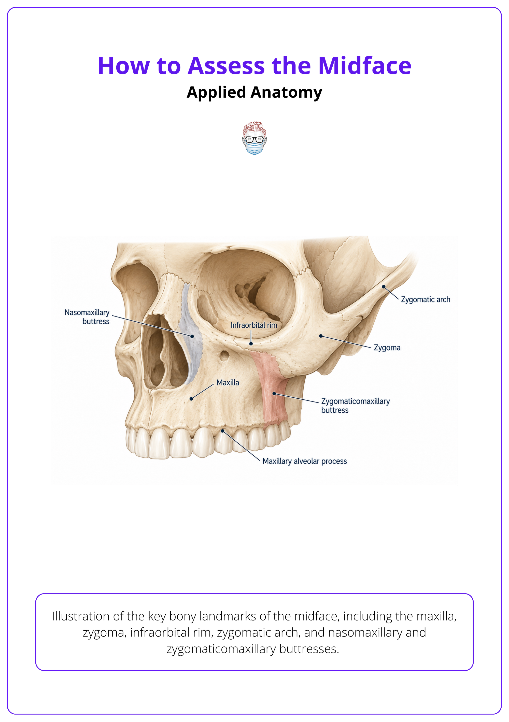
- Boundaries: lower orbital rim superiorly, zygomatic arch laterally, nasolabial fold medially, upper dentition inferiorly.
- Buttresses: assess nasomaxillary, zygomaticomaxillary, infraorbital, and maxillary alveolar support.
- Zygoma: compare malar eminence, arch width, lateral orbital rim, and cheek projection.
- Maxilla: distinguish maxillary retrusion from mandibular excess when a Class III profile is suspected 1.
Soft-Tissue Layers
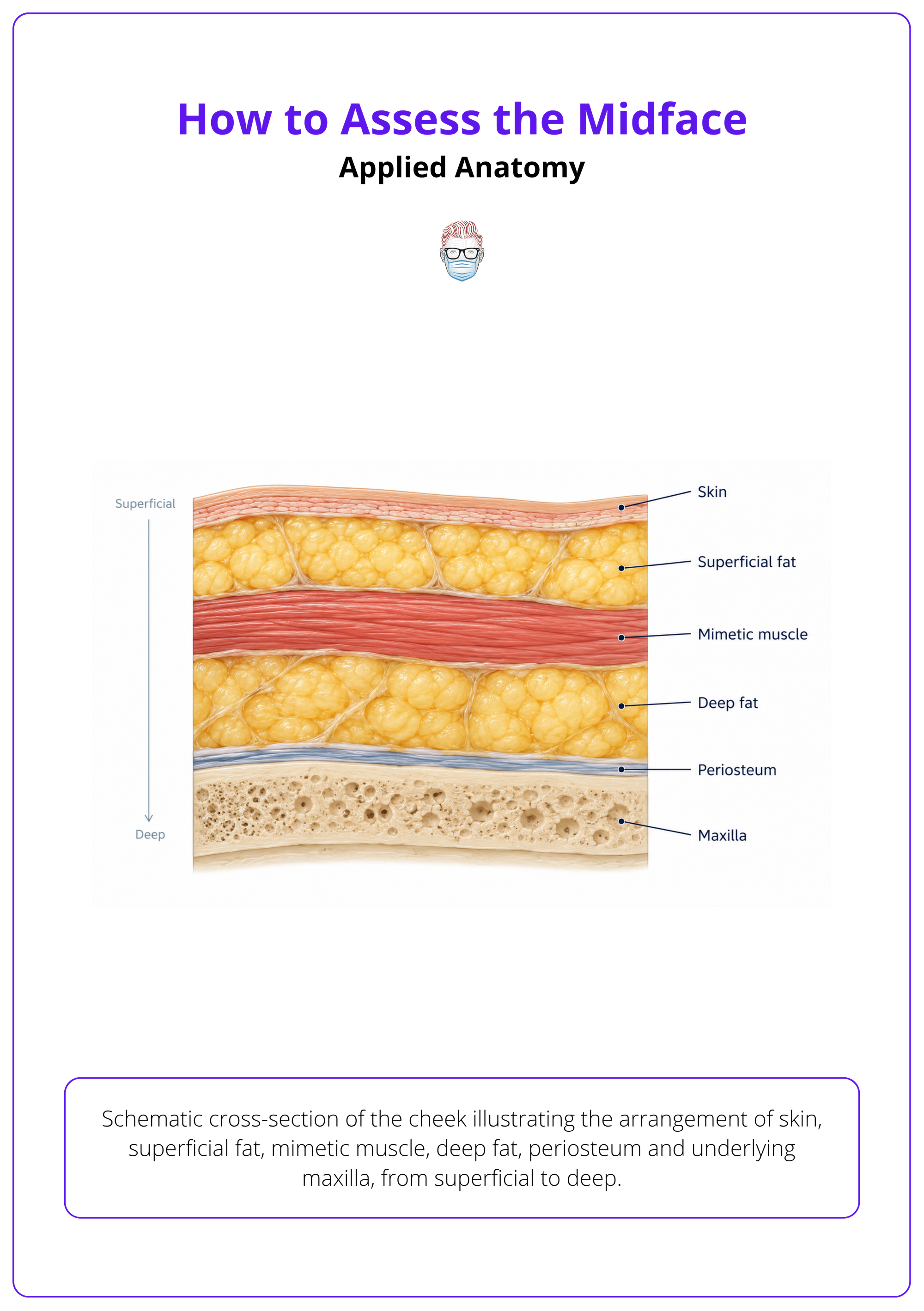
Analyse contour from deep support to mobile skin rather than attributing cheek shape to one structure.
- Layered assessment: evaluate skeleton, deep fat, muscle, superficial fat, and skin before planning intervention 2.
- Deep fat: loss of deep midface fat behaves like loss of structural support, not just a surface hollow 2.
- Retaining ligaments: orbitomalar, zygomatic, and masseteric cutaneous ligaments create tethering, folds, and descent patterns.
- Cheek fullness: separate buccal fat, masseter bulk, mandibular contour, and subcutaneous fat on assessment 3.
Neurovascular Anatomy
Map nerves and vessels because they change both diagnosis and procedural risk.
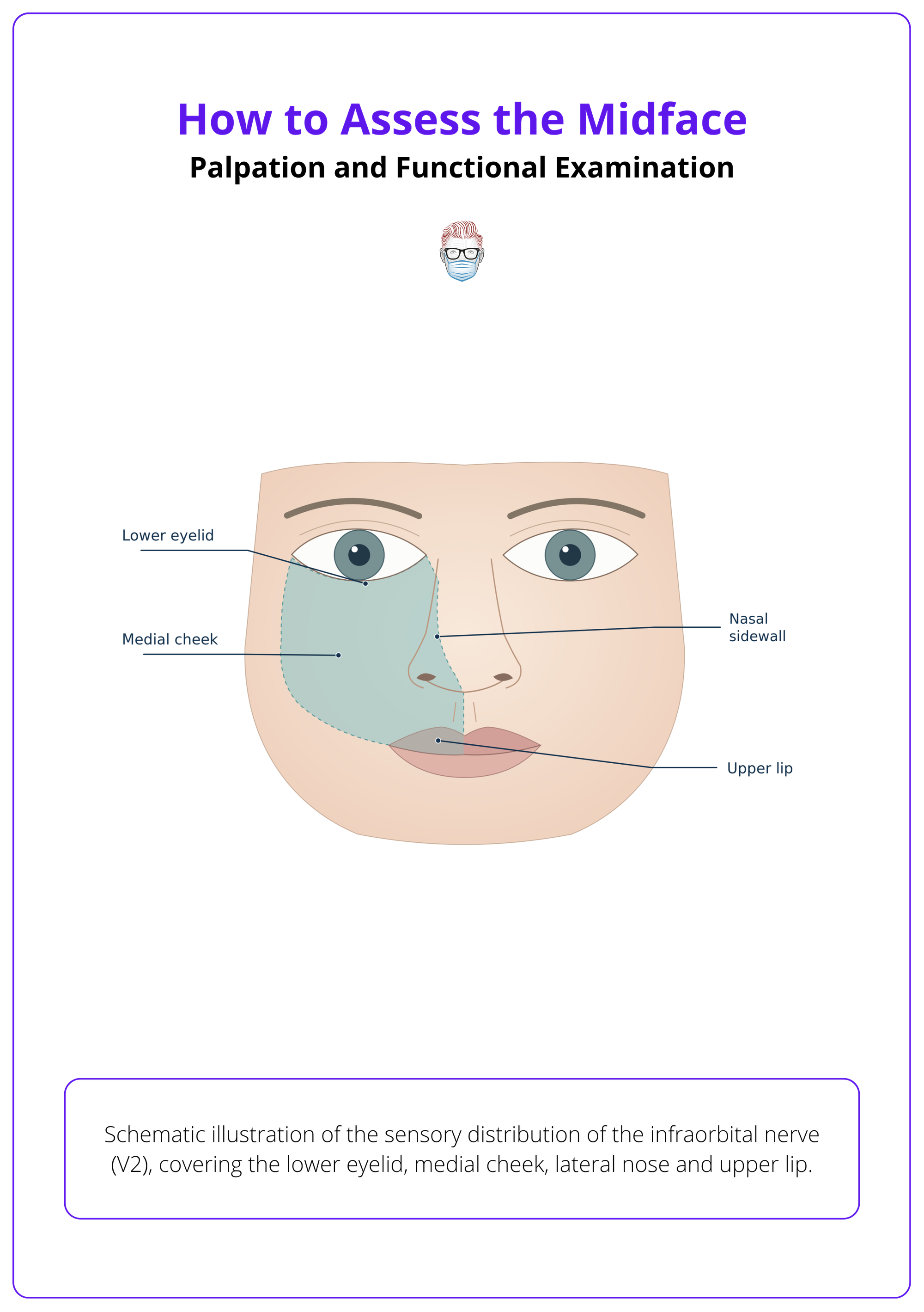
- Infraorbital nerve: test lower eyelid, nasal sidewall, upper lip, and maxillary dentition when involvement is possible 4.
- Trauma relevance: prior orbital floor or ZMC injury with infraorbital symptoms needs focused sensory follow-up 5.
- Facial nerve: screen zygomatic and buccal branch function through eye closure, smile, and upper lip animation.
- Vascular danger: a low-to-high approach tilted toward the infraorbital foramen risks arterial injury 2.
Assessment Goals
The assessment should define deformity type, asymmetry, skeletal support, soft-tissue volume and descent, orbital or occlusal involvement, functional risk, and clinical context.
A good midface assessment ends with a diagnosis, not a list of findings. Decide whether the problem is aesthetic, traumatic, congenital, developmental, oncologic, infective, or reconstructive.
Problem Definition
Classify the dominant abnormality before considering imaging or treatment.
- Skeletal problem: malar flattening, maxillary retrusion, buttress collapse, or facial-third disproportion.
- Soft-tissue problem: volume loss, cheek ptosis, lid-cheek hollowing, nasolabial deepening, or scarring.
- Functional problem: diplopia, malocclusion, sensory loss, pain, restricted mouth opening, or sinus symptoms.
- Integrated analysis: deformity planning should combine skeletal, dental, and soft-tissue findings 6.
Decision Consequences
Use the assessment to decide urgency, investigation, referral, and whether correction is safe.
- Treatment depth: determine whether the patient needs superficial correction, deep support, or a multilayer plan 2.
- Aesthetic goals: define goals, anatomy, expected improvement, and durability expectations before filler planning 7.
- Objective baseline: document severity in a way that supports indication and later outcome measurement 8.
- Safety anatomy: identify vascular danger before any midface correction, especially near the infraorbital foramen 2.
History and Context
History should identify mechanism, timing, congenital pattern, previous surgery or fillers, dental or sinus symptoms, ocular symptoms, sensory change, infection risk, oncologic history, and the patient’s stated concern.
The same cheek hollow can represent ageing, trauma, congenital hypoplasia, iatrogenic contour change, infection, or tumour recurrence. History makes the examination interpretable.
Trauma History
Mechanism and symptom timing determine whether subtle orbital, zygomatic, or nerve injury is being missed.
- Mechanism: ask about direct blow, assault, fall, road traffic injury, dental trauma, and previous fracture treatment.
- Orbital symptoms: document diplopia, pain on eye movement, blurred vision, enophthalmos, or globe displacement.
- Occlusal change: ask about altered bite, trismus, dental numbness, and maxillary mobility.
- Secondary deformity: untreated, delayed, or inadequately corrected ZMC trauma should raise concern for late midface deformity 9.
Developmental History
Congenital and orthodontic context prevents treating skeletal pathology as isolated cheek contour.
- Cleft history: record cleft type, hard palate closure timing, and orthodontic history because these affect later maxillary osteotomy need 10.
- Delayed closure: hard palate closure after 18 months in repaired cleft palate raises concern for greater maxillary hypoplasia 11.
- Syndromic pattern: Binder syndrome is a rare congenital midface malformation requiring syndrome-specific planning 12.
- Asymmetry history: ask whether asymmetry is lifelong, progressive, post-traumatic, or post-surgical.
Treatment History
Previous intervention changes anatomy, risk, and what the patient may reasonably expect.
- Facial surgery: record blepharoplasty, facelift, fracture fixation, orthognathic surgery, implants, fat grafting, and scars.
- Filler history: record product, timing, plane if known, complications, and reversibility; safety may differ between products 13.
- Vascular symptoms: ask about prior post-injection pain, blanching, skin necrosis, or visual symptoms because these map to rare serious filler risks 7.
- Ageing concern: clarify whether the complaint is malar projection, cheek fullness, or loss of the lower-lid-to-cheek ogee curve 2.
Inspection and Surface Analysis
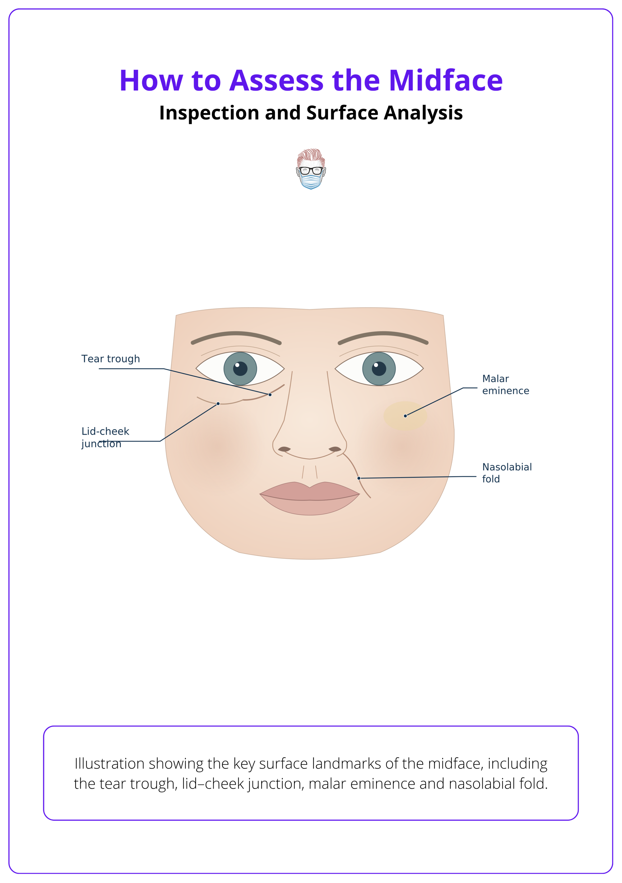
Visual assessment should identify symmetry, malar projection, lid-cheek contour, tear trough, nasojugal groove, nasolabial fold, cheek descent, swelling, scars, ocular position, dental show, and facial proportions.
Inspect before touching. Many midface diagnoses are pattern-recognition problems, but the pattern must be checked from several views.
Standard Views
Use fixed views so asymmetry is not created by head position or lighting.
- Frontal view: compare malar height, cheek width, orbital level, nasal deviation, dental midline, and facial thirds.
- Oblique views: assess malar highlight, tear trough depth, lid-cheek transition, and zygomatic projection.
- Lateral view: assess convexity, maxillary support, nasolabial angle, and lower facial balance.
- Basal view: use worm’s-eye or basal inspection for arch asymmetry, dental show, and maxillary cant.
Malar Projection
Compare projection side-to-side and against the orbit, nose, and upper lip.
- ZMC trauma: compare facial symmetry and malar projection because restoration targets these findings 14.
- Ageing pattern: look for malar flattening, nasolabial fold deepening, soft-tissue descent, and tired skeletal-volume appearance 7.
- Buccal fullness: strong malar support masked by excessive roundness may fit buccal fat contour assessment 3.
- Hypoplasia: a concave midface profile, anterior crossbite, or transverse arch deficiency suggests maxillary deficiency 10.
Lid-Cheek Unit
Separate lower eyelid pathology from true midface descent or volume loss.
- Lid-cheek junction: look specifically for concavity when assessing age-related midface change 2.
- Tear trough: record depth, lateral extension, eyelid bag relationship, and lower eyelid height 15.
- Age context: tear trough severity may increase with age, so compare appearance with age-appropriate baseline 15.
- Scleral show: inspect at rest and downgaze; asymmetry may reflect retraction, orbital change, or prior surgery.
Nasolabial-Cheek Unit
Identify whether folds and fullness come from descent, volume, scarring, or skeletal support loss.
- Nasojugal groove: document upper-cheek flattening and nasojugal prominence 2.
- Nasolabial fold: inferior collapse of nasolabial fat can accentuate both the fold and groove 2.
- Post-traumatic ptosis: record cheek and nasolabial ptosis because resuspension may restore preinjury aesthetics 16.
- Cheek fullness: do not attribute lower-cheek fullness to buccal fat alone without considering masseter, mandible, and subcutaneous fat 3.
Palpation and Functional Examination
Palpation and function should confirm bony steps, instability, tenderness, sinus involvement, infraorbital nerve deficit, facial nerve function, ocular motility, globe position, occlusion, mouth opening, and intraoral pathology.
Hands confirm what inspection suggests. Examine from orbit to dentition, then test sensation, animation, and function.
Skeletal Palpation
Palpate named buttresses so imaging requests and referrals are anatomically precise.
- Orbital rim: feel for step deformity, tenderness, mobility, crepitus, and rim malposition.
- Zygomatic arch: palpate laterally and intraorally if trismus suggests arch impingement.
- Maxillary buttresses: assess nasomaxillary and zygomaticomaxillary support for tenderness or instability.
- Imaging correlation: localise deformity to landmarks that can later be measured on CT when needed 17.
Sensory Examination
Map infraorbital sensation and pain because nerve injury can persist and may change follow-up.
- Territory: test lower eyelid, cheek, nasal sidewall, upper lip, gingiva, and maxillary teeth.
- Modality: use light touch, pinprick, two-point comparison, allodynia, and dysaesthesia mapping.
- Neurosensory deficit: suspected infraorbital nerve injury should be graded rather than described vaguely 18.
- Neuralgia history: after orbital floor trauma, actively ask about infraorbital neuralgia and causalgia 19.
Ocular Examination
Treat ocular findings as functional findings, not cosmetic observations.
- Vision screen: check acuity, pupils, colour desaturation if concerned, and urgent ophthalmology triggers.
- Motility: test diplopia in all gazes and pain on movement; record restriction pattern.
- Globe position: compare enophthalmos, hypoglobus, proptosis, dystopia, and vertical orbital asymmetry.
- Lower eyelid: document lagophthalmos, lid laxity, scleral show, and lid-cheek support.
Oral Examination
Occlusion and intraoral findings identify maxillary, dental, and sinus involvement.
- Occlusion: record open bite, crossbite, premature contact, dental midline shift, and patient-reported bite change.
- Mouth opening: measure interincisal distance and note pain, trismus, or arch impingement.
- Intraoral palpation: assess maxillary vestibule, buttress tenderness, gingival sensation, dental trauma, and mobility.
- Sinus signs: check maxillary sinus tenderness, purulence, oroantral communication, or recurrent infection.
Dynamic Function
Test animation because midface contour can change substantially during expression.
- Smile: compare zygomaticus function, cheek elevation, nasolabial movement, and upper dental show.
- Facial nerve: screen eye closure, cheek puff, smile, and upper lip elevation for zygomatic or buccal weakness.
- Dynamic contour: smiling can reveal irregularities that affect superficial correction planning 2.
- Objective tools: facial paralysis assessment should include objective midface movement where available 20.
Measurements and Documentation
Record midface findings with standardised photographs, reproducible measurements, sensory maps, occlusal notes, scar diagrams, swelling diagrams, and red-flag documentation.
Documentation should let another clinician understand the deformity without re-examining the patient. It should also allow serial comparison.
Clinical Photography
Use reproducible images before interpreting subtle asymmetry.
- Views: frontal repose, frontal animation, right and left oblique, lateral, basal, and close lower eyelid views.
- Technique: standardise lighting, focal length, head position, background, and distance.
- Aesthetic baseline: objective volumetric endpoints, follow-up timing, and standard photography or imaging support midface assessment 7.
- Severity record: standard photographs and validated scales can document visible midface change 8.
Linear Measurements
Measure features that change planning, not every possible landmark.
- References: record facial thirds, interpupillary line, intercanthal distance, occlusal cant, and dental midline.
- Projection: compare malar height, malar convexity, cheek width, and lid-cheek hollow side-to-side.
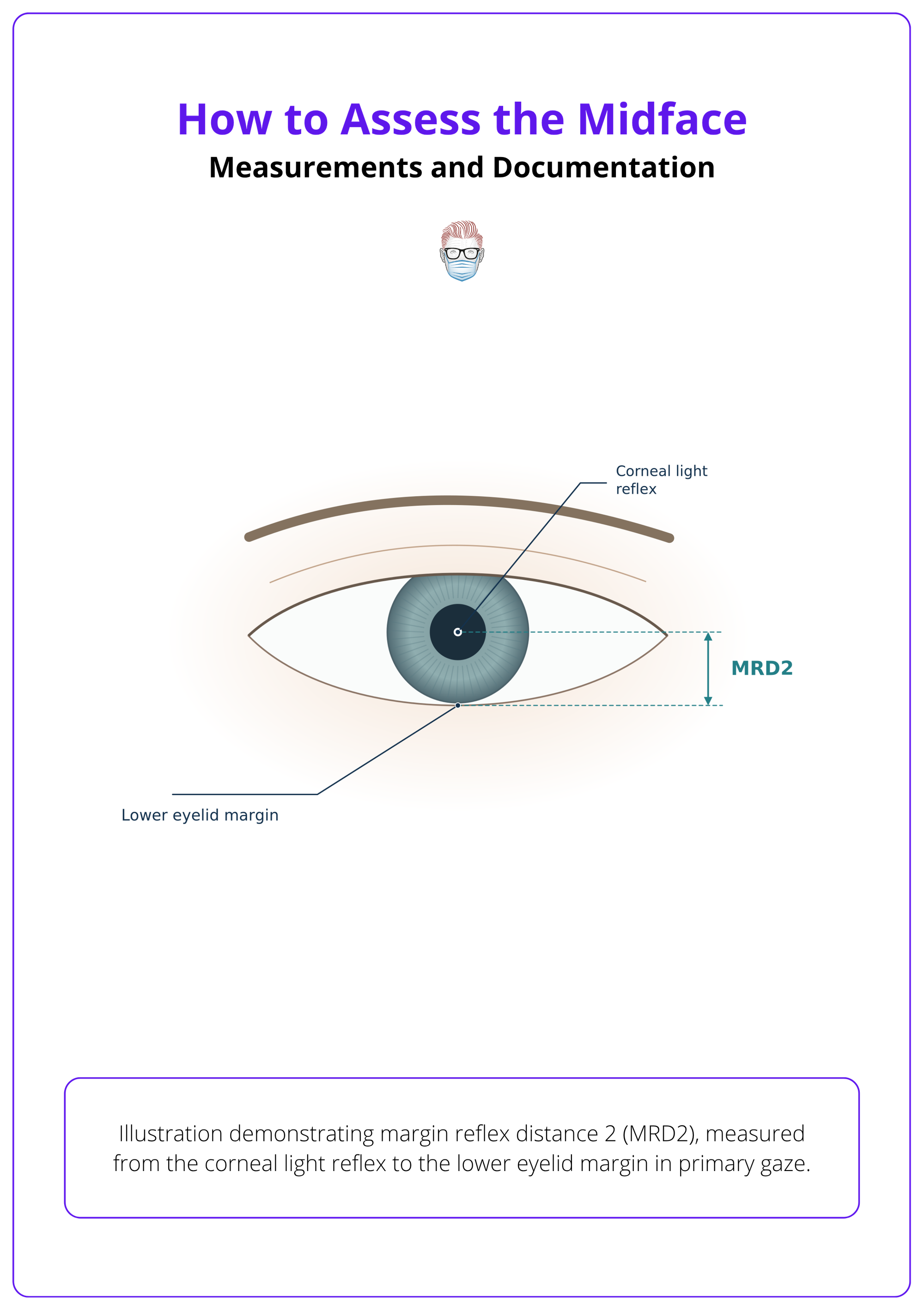
- Eyelid unit: document MRD2 and scleral show because static and dynamic lower eyelid findings guide treatment selection 21.
- Patient factors: record age and BMI when using anthropometric comparison because both can affect some 3D measurements 22.
Severity Scales
Use scales when they will guide treatment indication or serial follow-up.
- Indication: midface severity should be documented in a way that supports treatment indication and outcome measurement 8.
- Volume loss: a 5-point photonumeric scale can record midface volume loss more reproducibly than description alone 23.
- Merz Cheek Fullness Assessment Scale: the MCFAS provides reproducible clinician ratings of midface volume loss, with weighted kappa values greater than 0.6 23.
- Serial change: validated scales are preferable for follow-up because they improve consistency within and between raters 8.
Investigations and Imaging
Imaging is unnecessary for simple stable aesthetic assessment, but CT, orbital imaging, dental imaging, ultrasound, MRI, or specialist testing is indicated when trauma, orbital signs, skeletal discrepancy, mass, infection, implant, filler complication, or syndromic disease is suspected.
Investigations should answer a clinical question. Do not image reflexively, but do not let normal-looking skin override functional red flags.
No Imaging
Avoid imaging when the clinical problem is stable, superficial, and non-functional.
- Ageing assessment: stable volume loss, mild ptosis, or tear trough deformity may be assessed clinically and photographically.
- Mild asymmetry: longstanding unchanged asymmetry without pain, occlusal change, sensory deficit, or mass may not need imaging.
- Filler planning: imaging is not routine unless there is uncertainty about prior product, mass, infection, or vascular complication.
- Safety limit: no imaging does not mean no documentation, sensory examination, or ocular screen.
Computed Tomography
Use CT when bony, orbital, sinus, or post-traumatic anatomy will change urgency or planning.
- Facial bones CT: request for suspected ZMC, orbital floor, maxillary buttress, arch, or complex midface injury.
- Orbital CT: use when diplopia, motility restriction, globe displacement, enophthalmos, or rim step suggests orbital involvement.
- ZMC assessment: imaging is needed when clinical visualisation cannot define all zygomatic articulations.
- Landmarks: CT midface documentation may include zy-zy, zm-zm, n-pr, ns-pr, and ns-P distances when visible 17.
Adjunct Investigations
Choose adjunct imaging for the tissue, dental, or diagnostic question being asked.
- Cephalometry: use SNA angle and maxillary length measures when maxillary position or length is clinically relevant 1.
- CBCT: use in transverse maxillary deficiency planning when appliance or mini-implant position must be defined 24.
- Soft tissue: ultrasound may assess cheek volume, while MRI is preferred for masses, deep soft-tissue disease, or implant complications.
- Genetics: suspected syndromic midface hypoplasia, such as Miller syndrome, should prompt diagnostic confirmation 25.
Operative Verification
In complex fractures, imaging may be needed to verify reconstruction rather than just diagnose injury.
- Orbital reconstruction: endoscopic assessment and imaging can verify anatomic reconstruction in internal orbital fractures 26.
- Complex midface: intraoperative and postoperative imaging may be needed when restoring midface and internal orbital anatomy 16.
- Plain radiographs: do not rely on plain films to exclude clinically significant orbital floor or ZMC injury.
- Secondary deformity: major malar or orbital defects may require virtual planning, navigation, and updated imaging 9.
Assessment Synthesis and Red Flags
Convert findings into action: urgent referral for vision, entrapment, globe, infection, CSF leak, malocclusion, or progressive neurological signs; image structural disease; observe stable minor deformity; plan aesthetic or reconstructive care only after risk anatomy is defined.
The final assessment should state the likely diagnosis, severity, functional risk, missing information, and next step. Avoid vague conclusions such as “midface asymmetry for review.”
Emergency Referral
Escalate immediately when midface findings threaten vision, airway, occlusion, intracranial safety, or spreading infection.
- Vision change: reduced acuity, afferent pupillary defect, severe pain, or proptosis needs urgent ophthalmic input.
- Entrapment: diplopia with restricted motility, nausea, bradycardia, or severe pain suggests urgent orbital assessment.
- Globe displacement: hypoglobus, enophthalmos, or suspected orbital volume change after ZMC displacement is a functional red flag 14.
- Infection: rapidly progressive swelling, fever, dental sepsis, orbital cellulitis signs, or necrotic skin needs urgent escalation.
Decision Pathway
Match the next step to the dominant risk rather than the most visible deformity.
| Finding pattern | Next step | Rationale |
|---|---|---|
| Vision change or entrapment | Same-day orbital referral and CT | Function at risk |
| ZMC or orbital step with numbness | CT facial bones or orbit | Defines displacement |
| Malocclusion or maxillary mobility | Maxillofacial referral | Occlusion drives management |
| Stable ageing volume loss | Photography and aesthetic planning | No structural red flag |
| Progressive swelling or mass | Ultrasound or MRI pathway | Soft-tissue diagnosis |
- Observation: stable bruising, mild swelling, or longstanding asymmetry may be reviewed with photographs and safety-netting.
- Imaging threshold: lower the threshold when examination and mechanism disagree.
- Referral wording: include ocular findings, occlusion, sensation map, and exact bony tenderness.
Aesthetic Safety
Do not plan aesthetic correction until structural support, lower eyelid safety, and vascular risk are clear.
- Filler risk: assessment and consent before midface filler must address vascular occlusion, skin necrosis, and vision loss 7.
- Vascular anatomy: identify vascular variation and danger zones before injections because vascular injury is a key risk 2.
- Instrument path: avoid a low approach tilted toward the infraorbital foramen because of arterial injury risk 2.
- Buccal fat caution: hypoplastic malar bones are a red flag because excision can worsen contour by overcorrection 3.
Specialist Pathways
Use multidisciplinary input when the midface finding is congenital, syndromic, oncologic, occlusal, or reconstructive.
- Congenital hypoplasia: Binder syndrome requires syndrome-specific assessment rather than routine aesthetic planning 12.
- Uncertain plan: no single optimal surgical plan is established for Binder syndrome, so planning must be individualised 12.
- Syndromic clues: limb anomalies with midface hypoplasia should prompt assessment for Miller syndrome 25.
- Reconstructive planning: relate midface position and projection to the cranial base and mandible before orthognathic or reconstructive planning 6.
References
[1] Faria-Teixeira et al.. Craniofacial syndromes and class III phenotype: common genotype fingerprints? A scoping review and meta-analysis.. Pediatric research. 2024. doi:10.1038/s41390-023-02907-5
[2] Kaufman-Janette et al.. Injection Guidelines for Treating Midface Volume Deficiency With Hyaluronic Acid Fillers: The ATP Approach (Anatomy, Techniques, Products).. Aesthetic surgery journal. 2022. doi:10.1093/asj/sjac007
[3] Traboulsi-Garet et al.. Buccal fat pad excision for cheek refinement: A systematic review\.. Medicina oral, patologia oral y cirugia bucal. 2021. doi:10.4317/medoral.24335
[4] Meslemani et al.. Zygomaticomaxillary complex fractures.. Archives of facial plastic surgery. 2012. doi:10.1001/archfacial.2011.1415
[5] Devoti et al.. Characterization of Infraorbital Nerve Sequelae After Orbital Floor or Zygomaticomaxillary Complex Fractures.. The Journal of craniofacial surgery. 2022. doi:10.1097/SCS.0000000000007881
[6] Xia et al.. A New Clinical Protocol to Evaluate Cranio-maxillofacial Deformity and Plan Surgical Correction. Journal of Oral and Maxillofacial Surgery. 2009. doi:10.1016/j.joms.2009.04.057
[7] Safia et al.. Efficacy and Safety of Hyaluronic Acid Fillers for Midface Augmentation: A Systematic Review and Meta-Analysis.. Medicina (Kaunas, Lithuania). 2025. doi:10.3390/medicina61101823
[8] Carruthers et al.. Validated Assessment Scales for the Mid Face. Dermatologic Surgery. 2012. doi:10.1111/j.1524-4725.2011.02251.x
[9] Aman et al.. Secondary Reconstruction of the Zygomaticomaxillary Complex.. Seminars in plastic surgery. 2020. doi:10.1055/s-0040-1721761
[10] Tache et al.. The need for maxillary osteotomy after primary cleft surgery: A systematic review framing a retrospective study.. Journal of cranio-maxillo-facial surgery : official publication of the European Association for Cranio-Maxillo-Facial Surgery. 2020. doi:10.1016/j.jcms.2020.07.005
[11] Wlodarczyk et al.. A Meta-Analysis of Palatal Repair Timing.. The Journal of craniofacial surgery. 2021. doi:10.1097/SCS.0000000000007029
[12] Kerbrat et al.. Surgical Treatment for Patients With Binder Syndrome, Clinical Features and Associated Symptoms: A Systematic Review\.. The Journal of craniofacial surgery. 2022. doi:10.1097/SCS.0000000000008325
[13] Elrosasy et al.. Efficacy and safety of different hyaluronic acid fillers on cheek volume augmentation: systematic review and network meta-analysis.. Archives of dermatological research. 2024. doi:10.1007/s00403-024-03567-z
[14] Wang et al.. Contemporary Management of Zygomaticomaxillary Complex Fractures.. Seminars in plastic surgery. 2021. doi:10.1055/s-0041-1735812
[15] Yang et al.. A Quantitative Three-Dimensional Tear Trough Deformity Assessment and Its Application in Orbital Septum Fat Transposition.. Aesthetic plastic surgery. 2024. doi:10.1007/s00266-023-03474-0
[16] Mast et al.. Maxillofacial fractures: midface and internal orbit. Part 2: therapeutic options].. Der Unfallchirurg. 2012. doi:10.1007/s00113-011-2111-x
[17] Kulczyk et al.. The Association between Maxillary Sinus Dimensions and Midface Parameters during Human Postnatal Growth.. BioMed research international. 2018. doi:10.1155/2018/6391465
[18] Rath. Surgical treatment of maxillary nerve injuries. The infraorbital nerve.. Atlas of the oral and maxillofacial surgery clinics of North America. 2001. PMID:11665375
[19] Beigi et al.. Infraorbital Nerve Decompression for Infraorbital Neuralgia/Causalgia following Blowout Orbital Fractures: A Case Series.. Craniomaxillofacial trauma & reconstruction. 2020. doi:10.1055/s-0036-1592095
[20] Revenaugh et al.. Use of Objective Metrics in Dynamic Facial Reanimation: A Systematic Review\.. JAMA facial plastic surgery. 2018. doi:10.1001/jamafacial.2018.0398
[21] Rafizadeh et al.. Surgical Correction of Cicatricial Lower Eyelid Retraction: A Systematic Review\.. Seminars in ophthalmology. 2024. doi:10.1080/08820538.2023.2273850
[22] Adekunle et al.. Facial Anthropometry Measurements Using Three-Dimensional Stereophotogrammetry Analysis Among Nigerians.. The Journal of craniofacial surgery. 2022. doi:10.1097/SCS.0000000000008036
[23] Moradi et al.. Validation of a Midfacial Scale and Its Use in a Randomized, Evaluator-Blinded Study of CPM-HA-V.. Journal of drugs in dermatology : JDD. 2024. doi:10.36849/JDD.7981
[24] Brunetto et al.. Mini-implant assisted rapid palatal expansion (MARPE) effects on adult obstructive sleep apnea (OSA) and quality of life: a multi-center prospective controlled trial.. Progress in orthodontics. 2022. doi:10.1186/s40510-021-00397-x
[25] van et al.. Unveiling the Phenotypic Spectrum of Miller Syndrome: A Systematic Review\.. The Journal of craniofacial surgery. 2025. doi:10.1097/SCS.0000000000011501
[26] Mast et al.. Maxillofacial Fractures: Midface and Internal Orbit-Part II: Principles and Surgical Treatment.. Facial plastic surgery : FPS. 2015. doi:10.1055/s-0035-1563693
