

Nerve Injury
1. Peripheral nerves are groups of fascicles wrapped in an epineurium
2. Nerve injury results in chromatolysis & Wallerian degeneration
3. The degree of nerve injury is classified by the MRC Grading System
4. Type of injury is classified by Seddon or Sunderland Classifications
5. The epineurium is sutured in epineural nerve repairs.
Anatomy of a Nerve Injury
Key Point
A peripheral nerve is a group of fascicles surrounded by an epineurium. A fascicle is created from anatomical layers of functional and structural components.
Nerve anatomy consists of functional and structural components. The functional aspect is the nerve fibre and axons. The structural component is the supportive soft tissue anatomy - for example, the epineurium.
Neuron/Nerve Fibre Anatomy
The neuron is the nerve cell. It is the foundation of the peripheral nerve. There are many elements to its structure, the most important being the axon and its surrounding myelin and Schwann cells.
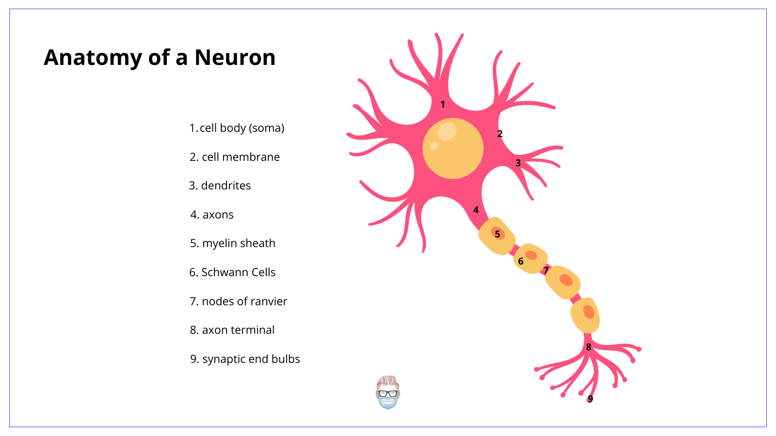
Functional components of the neuron include:
- Axons: nerve fibres that carry the impulses within the endoneurium.
- Schwann Cell: produce myelin sheath around myelinated nerves.
- Node of Ranvier: gaps between Schwann cells on myelinated nerves
- Saltatory conduction: nerve impulse jumping between nodes of Ranvier.
There are 3 different types of nerve fibres based on their myelin, diameter and velocity.
- Type A: large diameter, high velocity, myelinated
- Type B (pre-ganglionic): small diameter, low velocity, myelinated
- Type C (post-ganglionic): small diameter, small velocity, unmyelinated
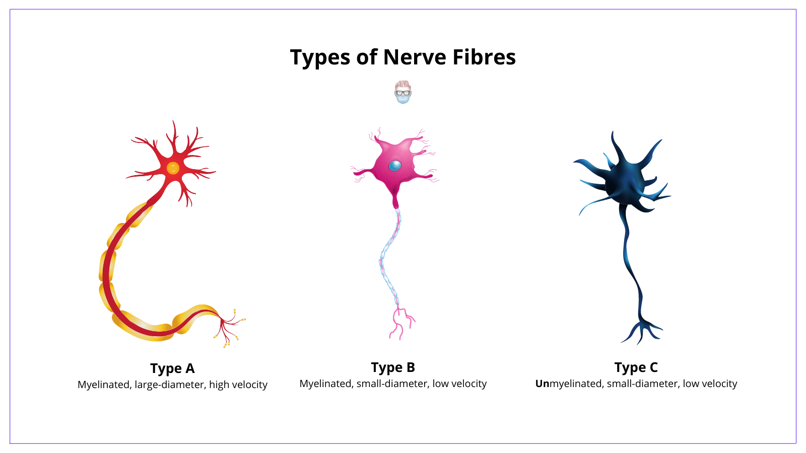
γ, δ based on their motor, proprioception and pressure functions.
Peripheral Nerve Anatomy
A peripheral nerve is a group of fascicles surrounded by an epineurium. Neurons (described above) help form the smallest unit of the peripheral nerve that can be surgically treated - fascicles. This is the foundation of a macroscopically visible peripheral nerve.
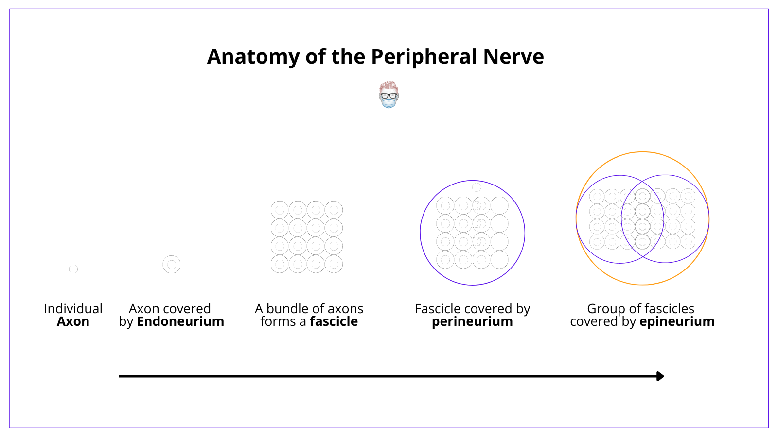
Structural components of the peripheral nerve include:
- Endoneurium: a collagen matrix surrounding an axon.
- Fascicle: a bundle of nerve fibres/axons wrapped in the perineurium.
- Perineurium: connective tissue of each fascicle creates a blood-nerve barrier.
- Inner Epineurium: surrounds the perineurium and contains blood vessels
- Outer Epineurium: surrounds groups of fascicles to form a peripheral nerve.
- Mesoneurium: adventitial layer assists in nerve gliding & epineural repairs.
Blood supply to the nerve can come in various ways. Most commonly:
- Arteriae nervosum: can help orientate your epineural nerve repair
- External vessels: supply the epineurium
- Capillary plexus: supplies the endoneurium.
Pathophysiology of a Nerve Injury
Key Point
Nerve injury results in chromatolysis in the proximal the injury, Wallerian degeneration distal to the injury and release of neurotrophic factors.
Wallerian Degeneration
Wallerian degeneration is the active degeneration of myelin and axons distal to the site of nerve injury. It was first described by Waller in 1850 and begins after a brief latent period.
Key steps in wallerian degeneration include:
- Increase in cytoplasmic calcium
- Proliferation of macrophages & Schwann cells (produce neutrophic factors).
- Degeneration and phagocytosis of axons and myelin.
- Formation of bands of bunger (endoneurial tubes to guide regenerating axons).

Chromatolysis
Chromatolysis is the process of proximal neuronal cell degeneration to the nearest node of Ranvier that can occur within several hours of injury.
Key steps in chromatolysis include:
- Degeneration to the nearest node of Ranvier.
- Axonal sprouts form a growth cone
- Growth cone provides contact guidance to find the correct distal stump.
NeuroTROPISM and NeuroTROPHISM
The direction of nerve regeneration is guided by neurotropism, neurotrophism and neurotropic factors.
- Neurotropism: selective, directional growth of fibres
- Neutrophism: non-selective, non-directional growth of fibres.
- Neurotrophic factors: release fromdistal target, produced by Schwann cells.
Examples of neurotrophic factors include nerve growth factor, insulin-like growth factor, epidermal growth factor (just to name a few!).
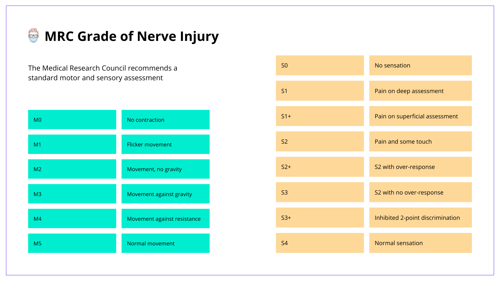
Assessment of a Nerve Injury
Key Point
The universal "language" for the grade of a nerve injury is published by the Medical Research council. It categories the motor or sensory injury based on clinical examination.
The assessment of a nerve injury should be based on a detail history and thorough clinical examination. The purpose of this section is to focus on key aspects specific to nerves.
MRC Grade of Nerve Injury
This is the official method for stratifying the degree of nerve injury based on clinical examination.
Additional Tests
There are a series of adjuncts to clinical examination that can help support your diagnosis. Some examples are list below.
- 2-point-discrimination: innervation density, 6mm static & 3mm dynamic.
- Monofilament: pressure thresholds (better for compression neuropathies)
- Electrophysiology Studies: Nerve conducton studies and EMGs.
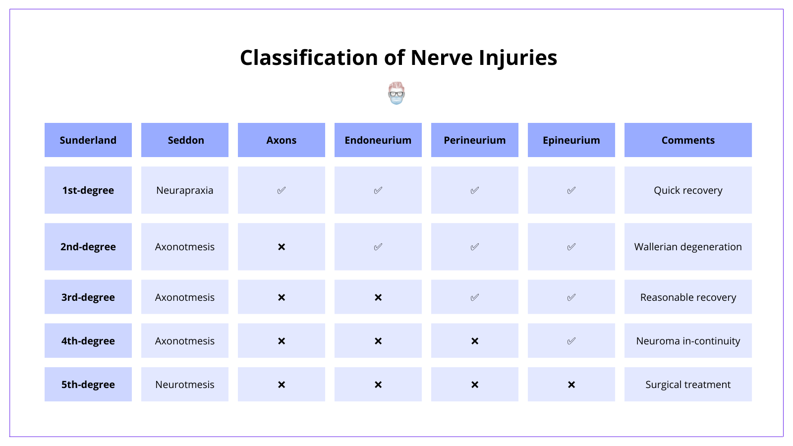
Classification of a Nerve Injury
Key Point
The Seddon and Sunderland classification systems are commonly used to describe the degree of nerve injury.
The classification of a nerve injury is by the Seddon or Sunderland classification. These classification systems are base on the type of injury to the nerve. They can guide management decisions and patient outcomes.
Flashcards
They are continually updated and are for thePlasticsPro users.
