Angiosomes

Angiosomes are a vascular territory. An underlying named artery supplies 3D composite blocks which are connected by choke vessels and can be used for tissue transfer.
Angiosomes

Summary on Angiosomes


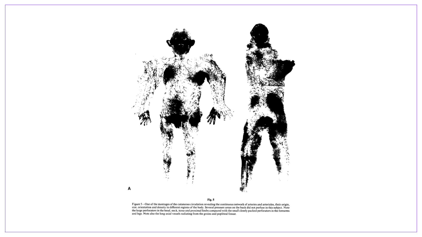
Definition of Angiosomes

An angiosome is a "vascular territory". It is a three-dimensional block of tissue supplied by a source vessel with its boundary outlined either by an anastomotic perimeter of reduced-calibre choke vessels or by true anastomoses with no reduction of vessel calibre.

The clinical significance of an angiosome is that it defines the tissues available for composite transfer. It helps our understanding of "zones of perfusion" and also the delay phenomenon.

💡
Tip: a dense network of vessels (for example, scalp, face, axillae, breasts, paraumbilical region, perineum, palms of the hand) correspond to the areas that sweat most profusely

A functional angiosome extends beyond the boundaries of an anatomical angiosome if it is connected by a true anastomosis instead of choke vessels. A choke vessel can be converted to a true anastomosis through the delay phenomenon.  

Original Taylor Publication in British Journal of Surgery

💡
Fun Fact: the first anatomical description of skin perfusion was by Carl Manchot in 1889. In 1936, Salmon used radiography to provide more detail. This was followed by the work of Taylor in 1987.


Principles of Angiosomes

The landmark 1987 publication by Palmar and Taylor has to lead to the development of specific angiosome principles.

  1. Angiosomes define the territory of the flap
  2. Source vessels follow the connective tissue framework of the body
  3. "Law of Equilibrium" - if one vessel is small, its adjacent or contralateral vessel will be larger. For example, a small ALT perforator is small will result in a larger perforator to TFL.
  4. Vessels travel from a fixed to a mobile area (long flaps can be raised from mobile areas)
  5. There is a neurovascular relationship (can raise with a nerve and vein).
  6. Vessels form a continuous unbroken network.
  7. A vessel's origin may change but its destination is constant.

💡
Tip: There is a close correlation between the angiosomes and the neurological dermatomes on the torso and in the head. 

Here is a video from one of Taylor's colleagues.

💡
Tip: a single vessel can supply multiple angiosomes. 

Example: DIEP/TRAM Flap

Hartrampfs Zone's of Perfusion provides a nice example of the angiosome theory. The abdomen is divided at the midline to form 2 hemi-abdomens. There are 4 zones and each zone has different levels of perfusion.  

The angiosome principle allows the abdomen to be divided into 4 zones of perfusion
Zones of Perfusion showing clinical use of angiosomes

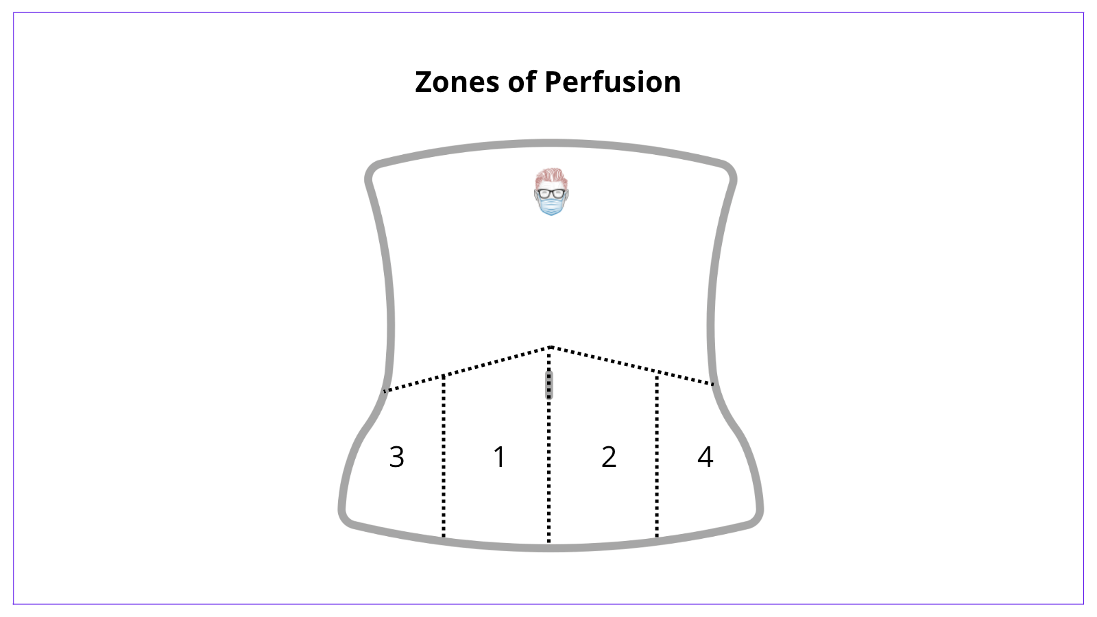
  1. Zone I always correlates to the zone of the selected perforator (most perfused)
  2. Zone II is adjacent to zone I on the contralateral hemi-abdomen.
  3. Zone III is lateral to zone I
  4. Zone IV is lateral to zone II (least perfused)

💡
Tip: The functional angiosome is the volume of tissue that clinically can be isolated on a source vessel. The area extends beyond its anatomical territory to capture an adjacent territory if connections are by choke anastomoses, or more if they are by true anastomoses.


Choke Vessels

Choke vessels can be defined as vessels that travel between two anatomical "vascular territories" or angiosomes.

Choke vessels are both arteries and veins. They connect two specific vascular territories. They are reorientated and dilated in the delay phenomenon. Their understanding plays an important role in plastic surgery.

Unlike choke vessels, a "true" anastomosis connecting two angiosomes will not decrease in size and are continuous. Choke vessels can be converted to "true" anastomosis via the delay phenomenon.  This is a permanent change that takes at minimum 3 days to occur.

Choke Vessels connect two angiosomes or anatomical territories
Choke Vessels

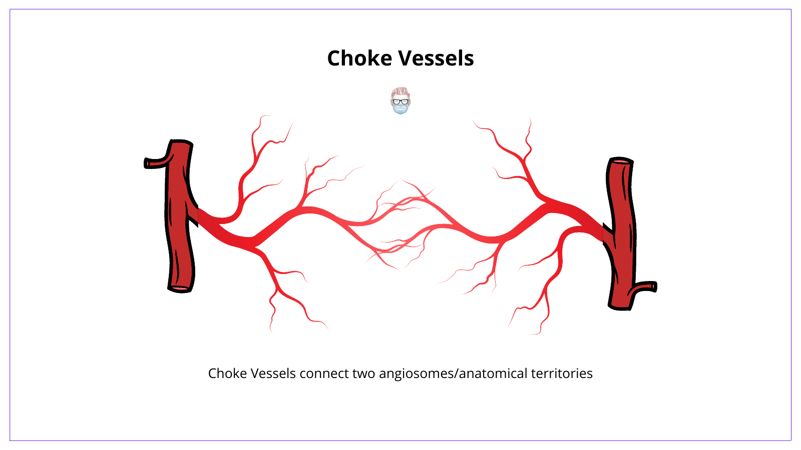
Choke Vessels have specific characteristics:

  • Arterial size and orientation are a product of growth
  • Arteries have a fixed destination but a varied origin.
  • Oscillating avalvular veins accompany choke arteries to allow free flow between valve channels and adjacent venous territories.

💡
Tip: superficial veins follow nerves, perforating veins follow perforating arteries. 


Flashcards on Angiosomes

This is for thePlasticsPro

Join the Club to enjoy unlimited access to all of thePlasticsFella.

Join the Club
Already have an account? Sign in

One platform for everything
Plastic Surgery

A curated suite of educational tools designed specifically for the evidence-based Plastic Surgeon.

Go Pro with a Free Trial

Great! You’ve successfully signed up.

Welcome back! You've successfully signed in.

You've successfully subscribed to thePlasticsFella.

Success! Check your email for magic link to sign-in.

Success! Your billing info has been updated.

Your billing was not updated.