

In this Article
5 Key Points on Brachial Plexus Anatomy
2. Segments of the plexus are: roots, trunks, divisions, cords, branches.
3. Course: intervertebral foramina to posterior triangle to the axilla.
4. Main branches: median, ulnar, radial, axillary, musculocutaneous.
5. Embryology: develops in the 5th week of gestation.

Brachial Plexus Anatomy & Embryology
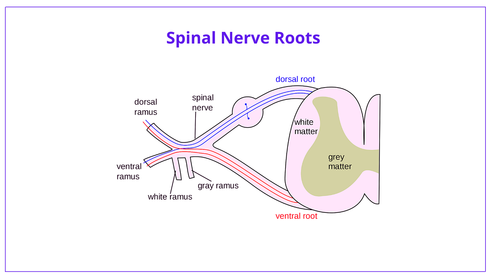
Motor nerve fibers:
- originate from cells within the basal plate of developing spinal cord.
- emerge to the ventral nerve root.
Sensory nerve fibers:
- originate from neural crest cells
- are found in the dorsal nerve root.
Brachial Plexus Anatomy Overview
Origins
The brachial plexus is formed by the anterior primary rami of spinal nerves C5, C6, C7, C8, T1.
Course
The plexus travels through the posterior triangle of the neck into the axilla, arm, forearm and hand.
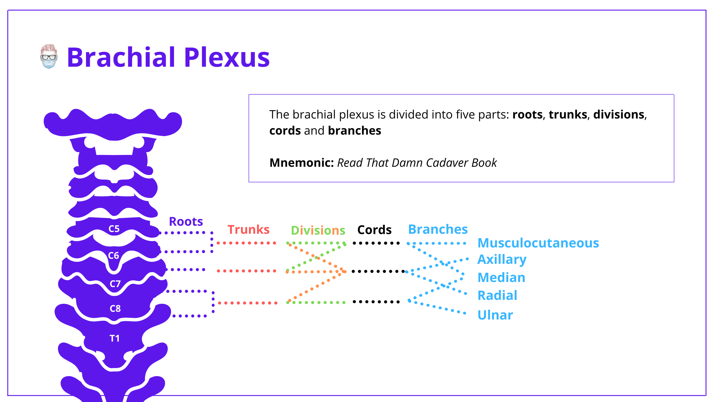
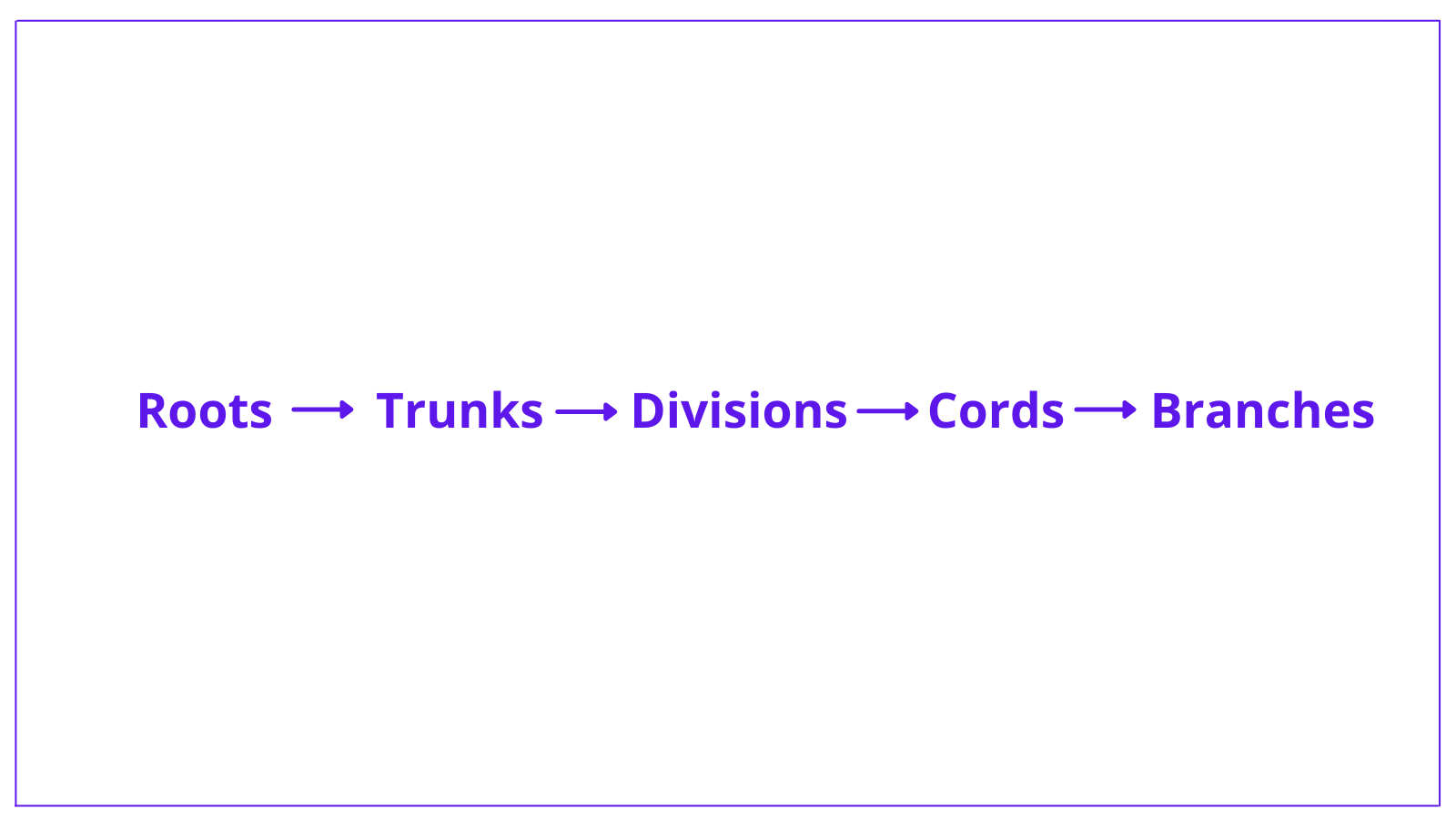
Structure
In general, the course followed by the intricate network is: Roots Trunks Divisions Cord Branches. The mnemonic is Read That Damn Cadaver Book.
Brachial Plexus Anatomy Roots
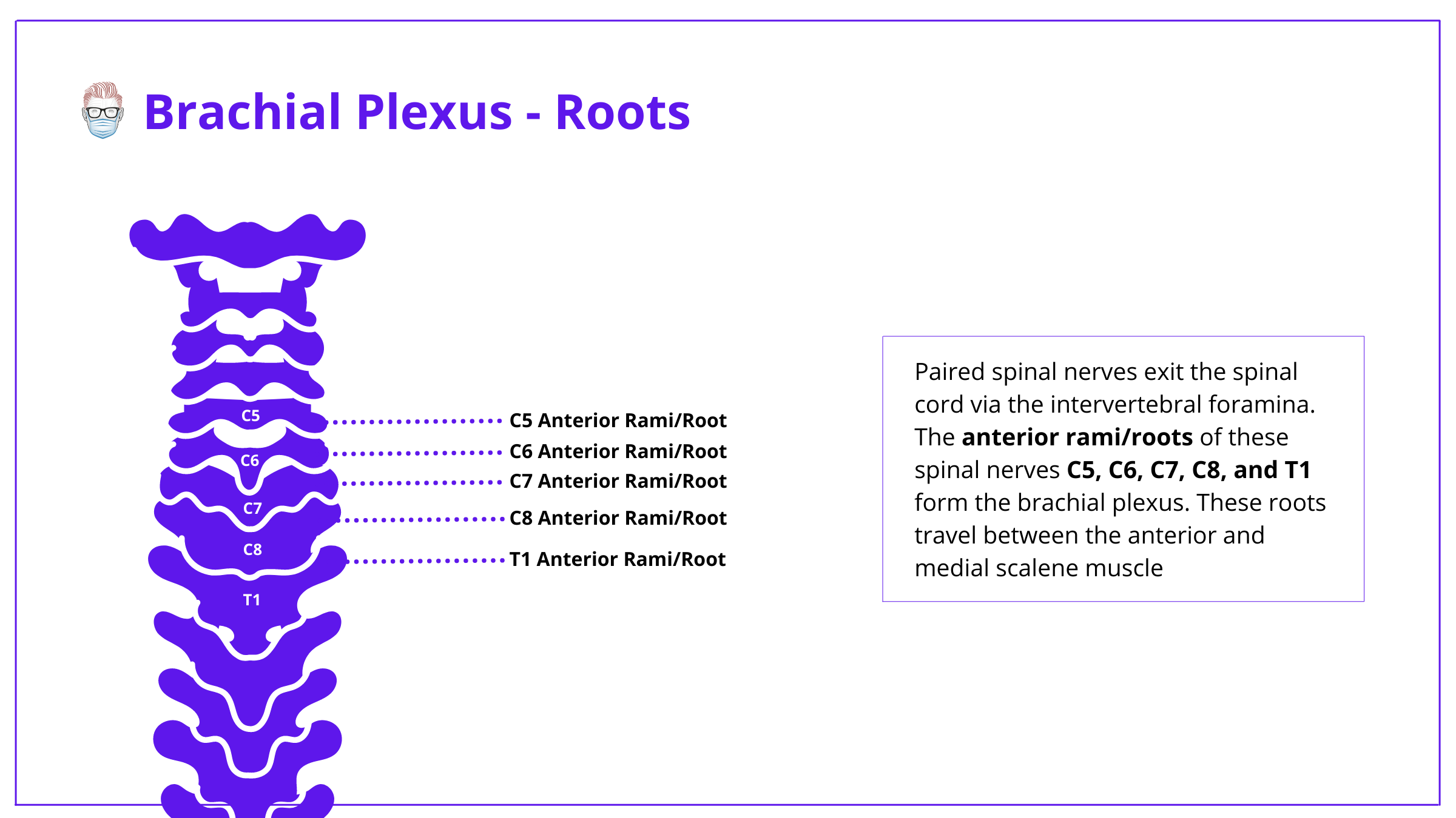
Brachial Plexus ‘roots’ refer to the anterior/ventral rami of spinal nerves C5 - T1. The dorsal rami innervate the skin and musculature of the intrinsic back muscles
Here are key points on the anatomy:
- Paired spinal nerves arise leave the spinal cord via the intervertebral foramina of the vertebral column and divide into an anterior and posterior ramus.
- The anterior rami form the roots of the brachial plexus whlst the posterior rami supply innervation to posterior skin and back muscles
- The roots travel between the anterior and medial scale towards the base of the neck.
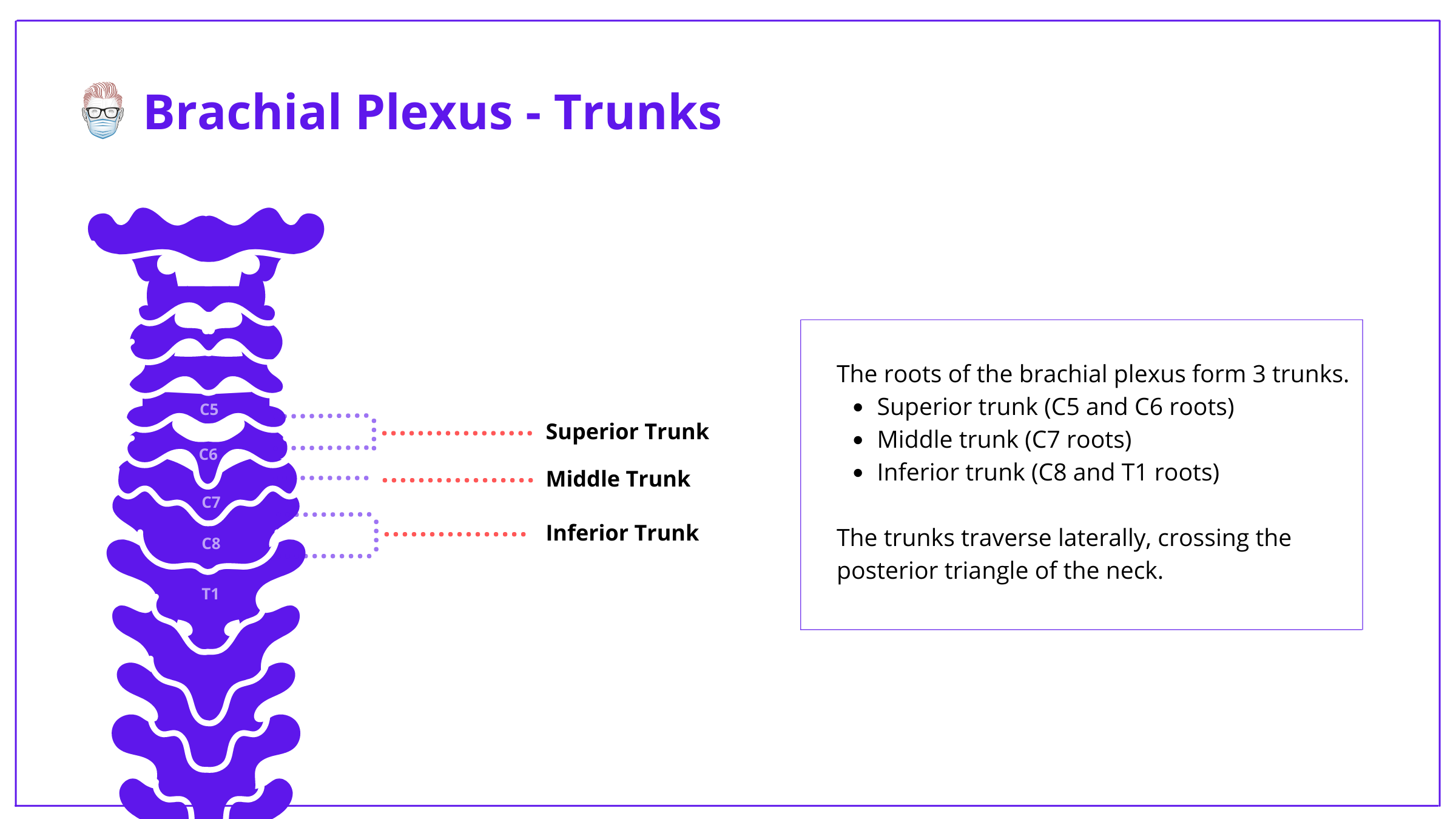
Brachial Plexus Anatomy Trunks
The C5-T1 brachial plexus roots form three trunks as they travel across the posterior triangle of the neck.
- Superior trunk – C5 and C6 roots.
- Middle trunk – C7 root.
- Inferior trunk – C8 and T1 roots.
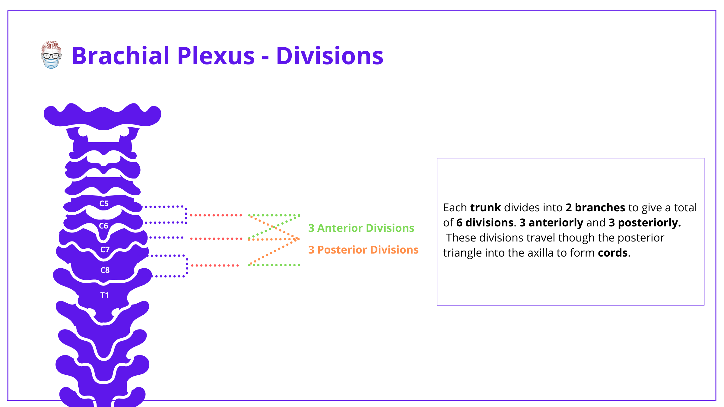
Brachial Plexus Anatomy Divisions
As the brachial plexus trunks exist the posterior triangle into the axilla, they divides into anterior (towards the front of the body) and posterior (towards the back of the body) divisions.
This results in the formation of 6 divisions:
- 3 anterior divisions
- 3 posterior divisions
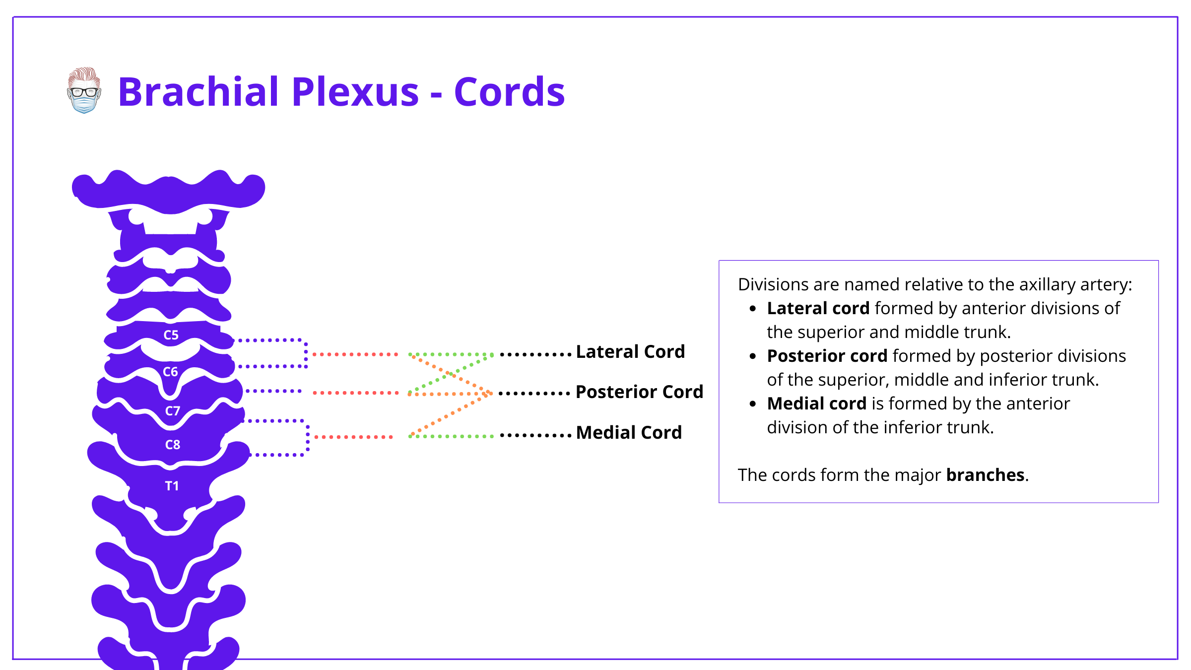
Brachial Plexus Anatomy Cords
Cords are formed from divisions and are named based on their relationship to the axillary artery.
- Lateral cord (C5-C7): Anterior divisions of Superior and Middle trunks
- Medial Cord (C8, T1): Anterior division of the Lower trunk.
- Posterior cord (C5-T1): Posterior divisons of the three trunks.
Brachial Plexus Anatomy Branches
The brachial plexus is the origin of a large number of nerve branches. Theres can be classified as "main branches" and "minor branches".
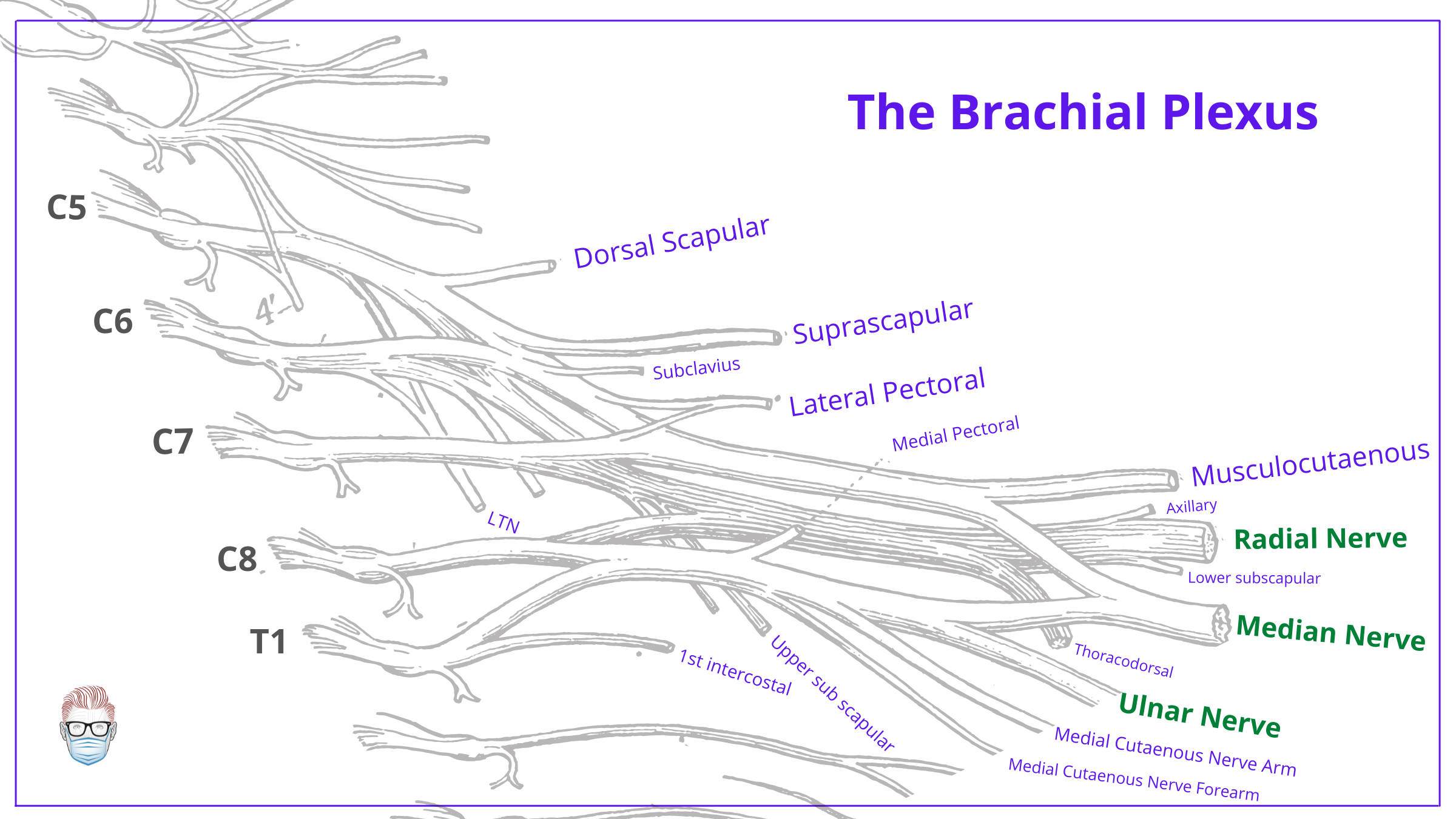
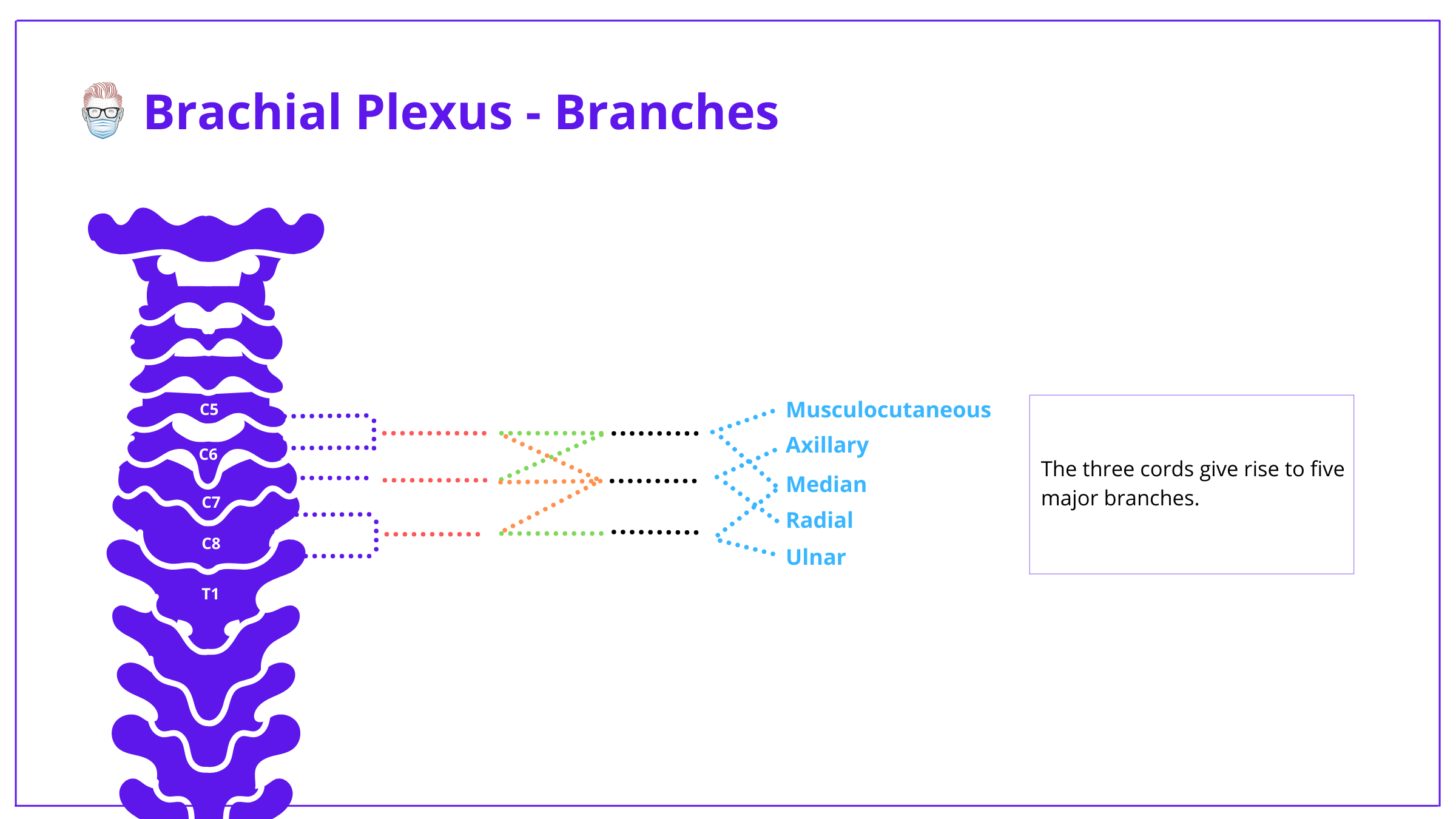
Main Branches of Brachial Plexus
The 5 main branches that originate from the brachial plexus are:
- Axillary Nerve: C5 and C6.
- Musculocutaneous nerve: C5, C6, C7.
- Radial Nerve: C5, C6, C7, C8, T1
- Median Nerve: C6, C7, C8, T1
- Ulnar Nerve: C8 and T1.
These main anatomical branches are seen in the image below.
Minor Branches of Brachial Plexus
Minor branches arise from multiple locations on the brachial plexus - from the root to the terminal divisions.
These minor branches can be classified as supraclavicular and infraclavicular. A simpler way is to catergorised them from their origin:
- Roots: Dorsal scapular (C5), Long thoracic (C5-7)
- Upper Trunk: Suprascapular (C5-C6), subclavius (C5-C6)
- Lateral Cord: Lateral pectoral (C5-7)
- Medial Cord: Medial pectoral, medial cutaneous of arm & forearm (C8,T1)
- Posterior Cord: Upper & lower subscapular (C5,6), thoracodorsal (C6-8)
Here is a table showing innervation and function of the minor branches.

Brachial Plexus Anatomy Landmarks
The brachial plexus originates in the posterior triangle of the neck.
- It extends from the lateral border of the scalene anterior muscle to the caudal (inferior) border of the pectoralis minor where each of the three cords divides into two terminal branches in the axillary fossa.
- The posterior triangle is covered superficially by the skin and superficial fascia, then by the platysma muscle and deep fascia. It is pierced by supraclavicular branches of the cervical plexus (C3 and C4).
- At the base of the neck, the plexus is behind the clavicle and subclavius muscle.
- Transverse cervical and suprascapular arteries cross over parts of the plexus
There are 2 methods to identify the brachial plexus: clinically and surgically.
On Palpation
The brachial plexus can be felt by running the fingers along the superior border of the clavicle, at the base of the neck. At about halfway, the cable can be felt running diagonally.
Surgically
While dissecting the upper limb, identify the characteristic ‘M’ pattern formation (Musculocutaneous, Median and Ulnar Nerves), usually superficial to the axillary artery. One can work backwards to identify the cords, divisions and branches.
Different ways to learn Brachial Plexus Anatomy
There are so many different ways to learn the anatomy of the brachial plexus. Here are 3 different ways you might find useful.
Let me introduce you to this amazing structure. It scares many people who learned it as medical or paramedical student, but to me it’s wonderful clever & beautiful. A triumph of developmental neurology and essential in all we do- let me introduce the brachial plexus via a thread pic.twitter.com/2WhIAAblEF
— Tom Quick (@TJQPNI) May 3, 2020
this.
— thePlasticsFella (@PlasticsFella) February 10, 2022
by @alexludai 🚂 pic.twitter.com/NeMqQWNRuV
References on Brachial Plexus Anatomy
- Bayot ML, Nassereddin A, Varacallo M. Anatomy, Shoulder and Upper Limb, Brachial Plexus. [Updated 2020 Jul 27]. In: StatPearls [Internet]. Treasure Island (FL): StatPearls Publishing; 2021 Jan-
- Warade AC,Jha AK,Pattankar S,Desai K, Radiation-induced brachial plexus neuropathy: A review. Neurology India. 2019 Jan-Feb;
- Abdullah S, Bowden RE. The blood supply of the brachial plexus. Proc R Soc Med. 1960 Mar;53:203-5.
- Desai SS,Varacallo M, Anatomy, Shoulder and Upper Limb, Musculocutaneous Nerve 2018 Jan.
- Elzanie A,Varacallo M, Anatomy, Shoulder and Upper Limb, Deltoid Muscle 2018 Jan.
- Pester JM,Varacallo M, Median Nerve Block Techniques 2018 Jan
- Gragossian A,Varacallo M, Radial Nerve Injury 2018 Jan.
- Pester JM,Varacallo M, Ulnar Nerve Block Techniques 2018 Jan.
- Thatte MR, Mehta R. Obstetric brachial plexus injury. Indian J Plast Surg. 2011;44(3):380-389. doi:10.4103/0970-0358.90805
- Kuhn JE, Lebus V GF, Bible JE. Thoracic outlet syndrome. J Am Acad Orthop Surg. 2015 Apr;23(4):222-32.
- Johnson D. Pectoral girdle and upper limb: overview and surface anatomy. Standring S,et al, ed-in-chief. In:Gray’s Anatomy: The Anatomical Basis of Clinical Practice. 40th ed. Churchill Livingstone Elsevier; 2008:777-790: chap 45, sec 6: Pectoral Girdle and Upper Limb.
- Shah HR, Takwale AB, Thatte MR. A Brachial Plexus with an Aberrant Middle Trunk: A Rare Anomaly. Indian J Plast Surg. 2019;52(3):364-365. doi:10.1055/s-0039-3401523
