In this Article
- 5 Key Points
- Overview of PIPJ Anatomy
- PIPJ Movement
- Articular Surfaces of PIPJ
- PIPJ Collateral Ligaments
- PIPJ Volar Plate
5 Key Points
- Joint: PIPJ is a bicondylar, synovial, hinge joint between the articular surfaces of the middle and proximal phalanx.
- Structure: a 3-sided box created by the volar plate and collateral ligaments.
- Collateral ligaments: resist against radial and ulnar deviation
- Volar plate: primary role in preventing hyperextension of the joint
- PIPJ dislocation: 2 sides of the box must be disrupted.
Overview of PIPJ Anatomy
The proximal interphalangeal joint (PIPJ) is a bicondylar, synovial, hinge joint. The articular surfaces of the proximal and middle phalanx are stabile in saggital movements (flexion and extension) but have limited tolerance to angular, axial, and rotational stress.
It is best visualized as a 3D structure, in which the articular surfaces of the middle and proximal phalanx are wrapped in a 3-sided-box.
This box provides stability to the joint and is formed by:
- Floor: volar plate and volar lip of the middle phalanx.
- Walls: collateral ligaments.
- Roof: joint capsule and extensor apparatus.
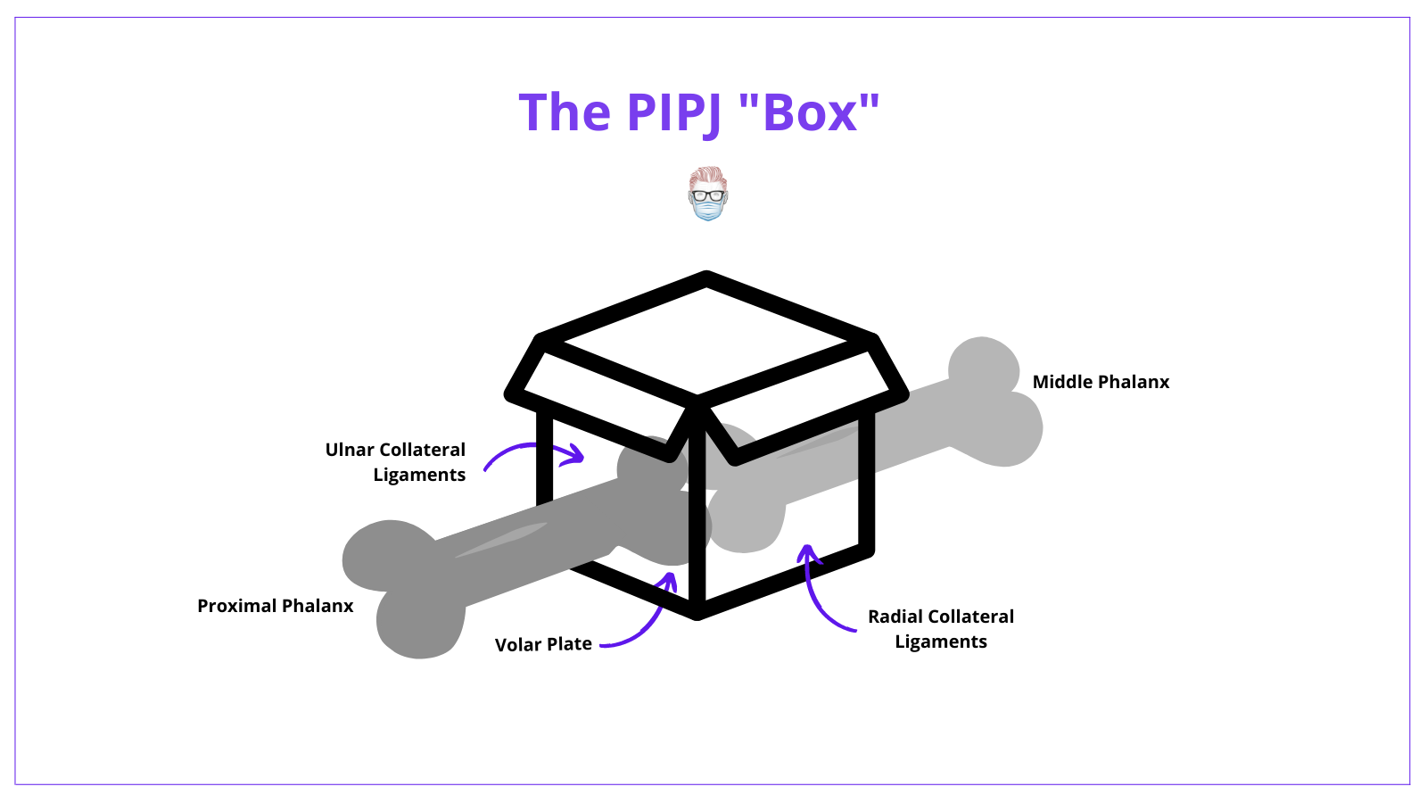
In a PIPJ dislocation, at least 2 sides of this box must be affected. Typically, collateral ligaments fail proximally while the volar plate avulses distally.
It is used as a landmark when describing the dorsal/extensor zones of the Hand. PIPJ represents "zone 3".
PIPJ Movement
There are two main active forces on the PIPJ:
- Extension: via central slip inserting on the dorsum of the middle phalanx.
- Flexion: via FDS tendon inserting on the volar aspect of the middle phalanx.
These forces provide the PIPJ with a motion arc 0° to 120°. This provides ~85% of the total finger motion required in functional grip.
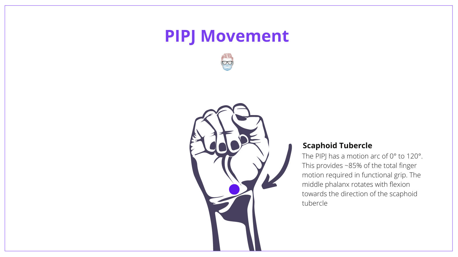
It is worth noting the unique anatomy of the PIPJ. The index and middle fingers have prominent ulnar condyles, whilst the ring and little fingers have prominent radial condyles. This results in the middle phalanx rotating with flexion towards the direction of the scaphoid tubercle.
Articular Surfaces of the PIPJ
The proximal interphalangeal joint (PIPJ) articular surfaces are the head of the proximal phalanx and base of the middle phalanx. Key anatomy features are described below.
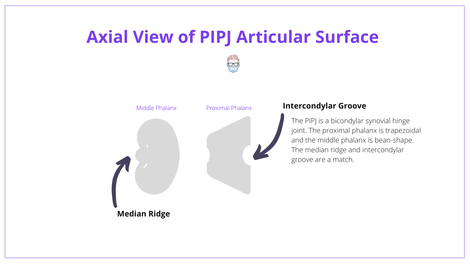
Head of the Proximal Phalanx
- Trapezoidal shape
- Intercondylar groove (this fits into the median ridge of the middle phalanx)
- Palmar tilt of ~20 degrees.
Base of Middle Phalanx
- A 'bean-shaped' structure separated by a median ridge
- The Median Ridge sets into the intercondylar groove of the proximal phalanx, like a tongue-in-groove. This provides resistance to axial rotation and translation forces acting on joints.
- The volar lip is an important stabilizing structure.
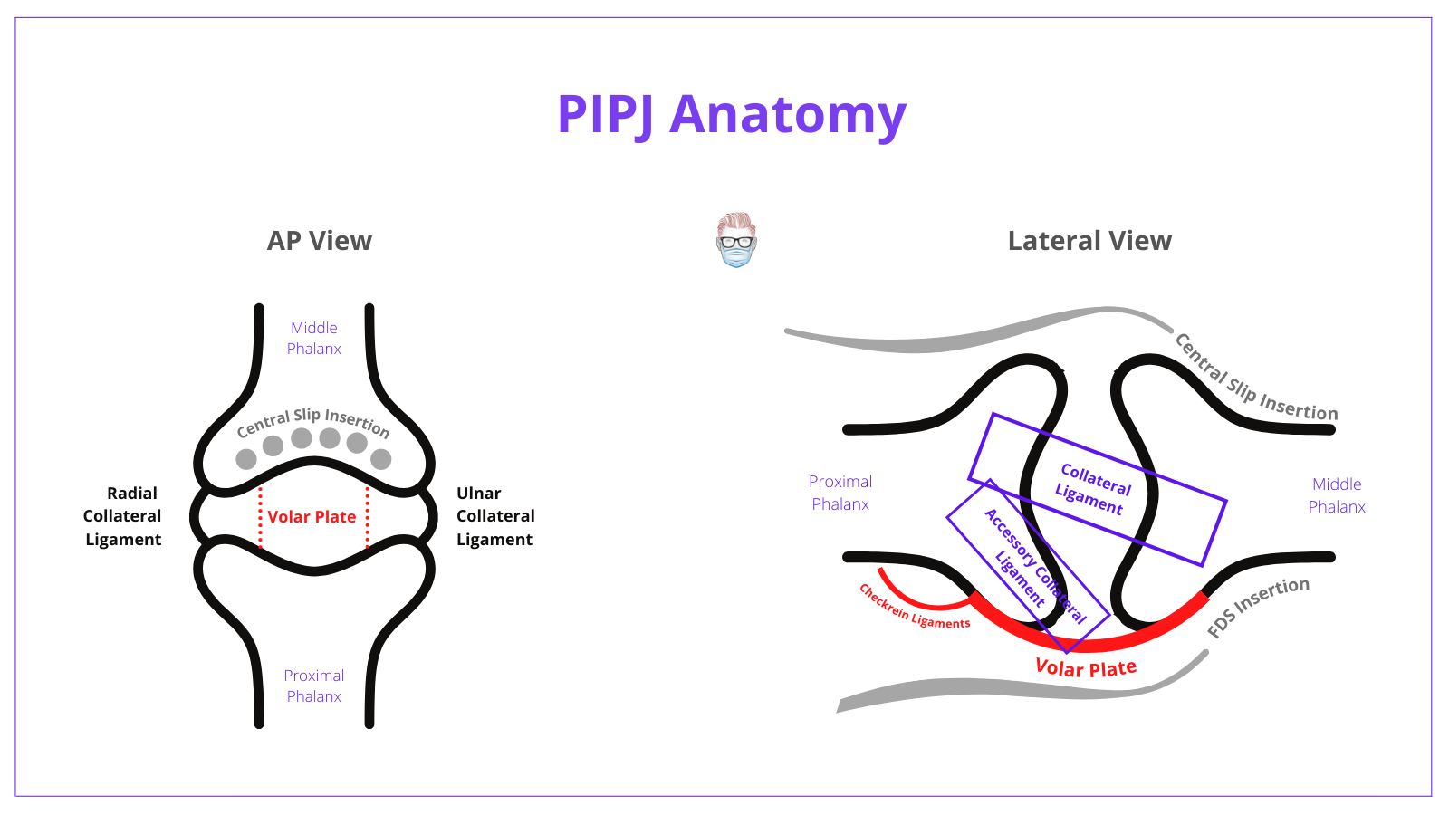
PIPJ Collateral Ligaments
The collateral ligaments of the proximal interphalangeal joint (PIPJ) are stabilizing anatomical features that resist against ulnar and radial deviation.
There are 2 types of collateral ligaments - accessory & proper collateral ligaments. They have a common origin and are often distinguishable only by their insertion points:
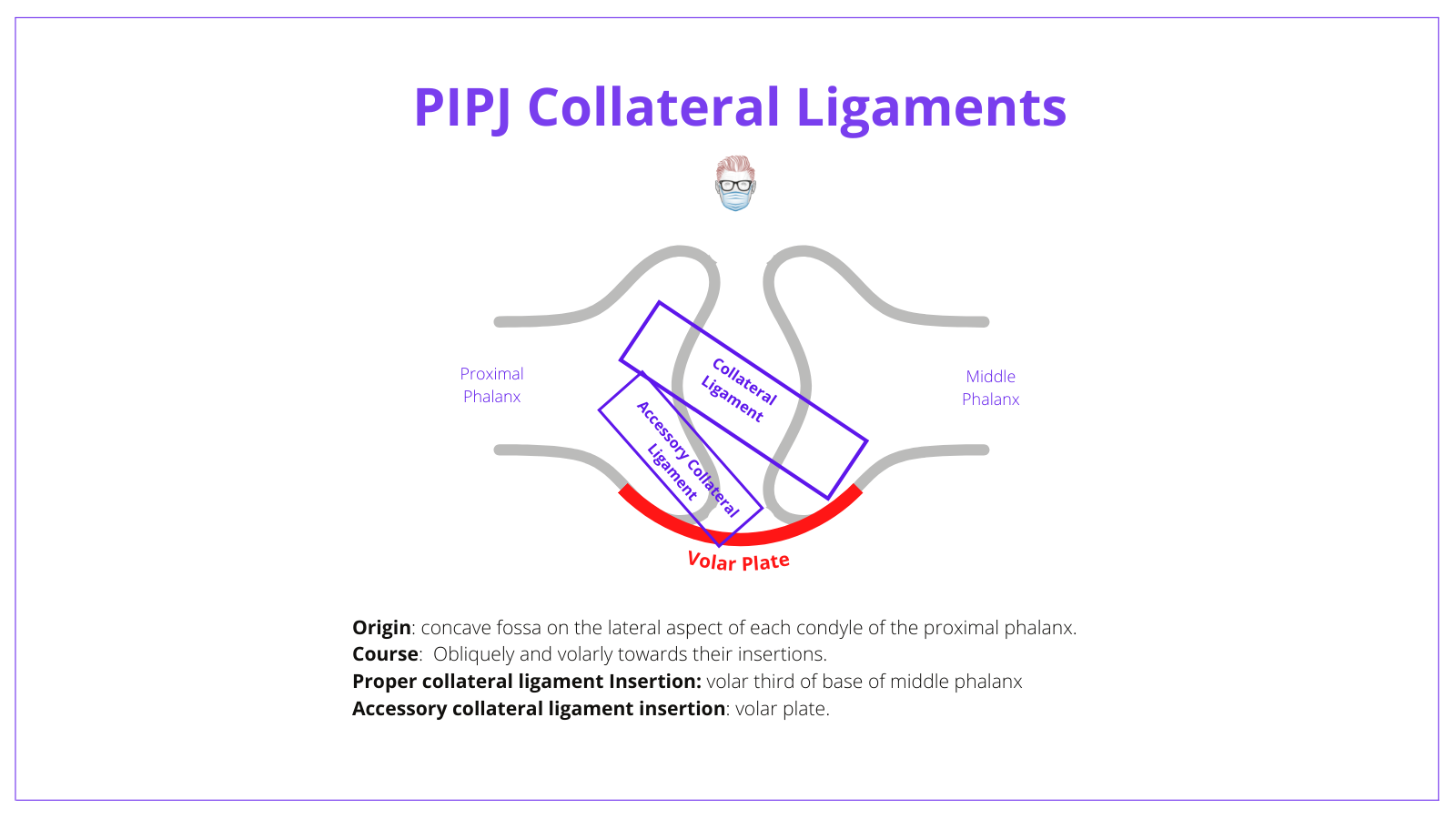
Anatomy
Key anatomical points for PIPJ collateral ligaments are:
- Origin: a common origin at the proximal phalanx
- Location: travel obliquely and volar to their insertions.
- Insertion: proper collateral ligament inserts onto volar base of the middle phalanx and the accessory collateral ligament inserts onto the volar plate.
The origin and insertion of the PIPJ collateral ligaments are through Sharpey's Fibers.
Function
The proper collateral ligments of the PIPJ have dorsal and the volar parts that act as a team during the full range of movement of the joint:
- During flexion: dorsal fibers become taut
- During extension: volar fibers become taut.
The accessory collateral ligaments of the PIPJ are taut in extension and relax on flexion. Their role is to:
- Suspend and stabilize volar plate
- Complete the joint capsule and act as a base for synovial lining
PIPJ Volar Plate
The volar plate is a fibrocartilaginous structure that forms the floor of the proximal interphalangeal joint (PIPJ). The insertion points of the volar plate are:
- Laterally: collateral ligaments
- Distally: volar base of the middle phalanx (mainly lateral margins)
It has a few unique anatomical features:
- Distally: dense fibrocartilaginous structure.
- Centrally: thin and confluent with volar periosteum of middle phalanx.
- Proximally: lateral aspects of volar plate form a pair of checkrein ligaments.
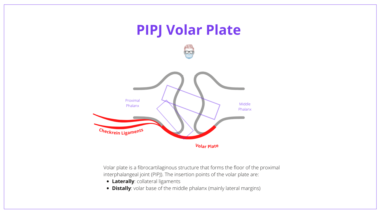
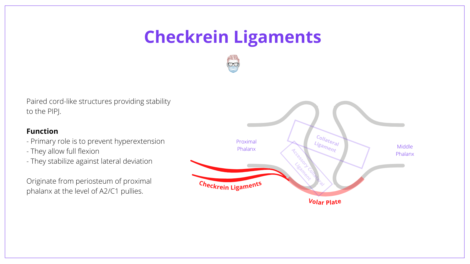
Checkrein ligaments
Checkrein ligaments are paired cord-like structures that provide further stability to the proximal interphalangeal joint (PIPJ).
Important points on their anatomy are:
- The primary role is to prevent hyperextension
- They allow full flexion
- They stabilize against lateral deviation if collateral ligaments are injured.
- Originate from periosteum of proximal phalanx at the level of A2/C1 pullies.