

Summary Card
Overview of Upper Limb Embryology
Upper limb development occurs during weeks 4-8 of gestation from the mesoderm and ectoderm.
Developmental Milestones
The limb bud appears on day 26 and differentiates into a limb by day 58.
Mechanisms of Development
Upper limb embryogenesis is based on 3 axes, 3 signalling zones, and 3 proteins.
Overview of Upper Limb Embryology
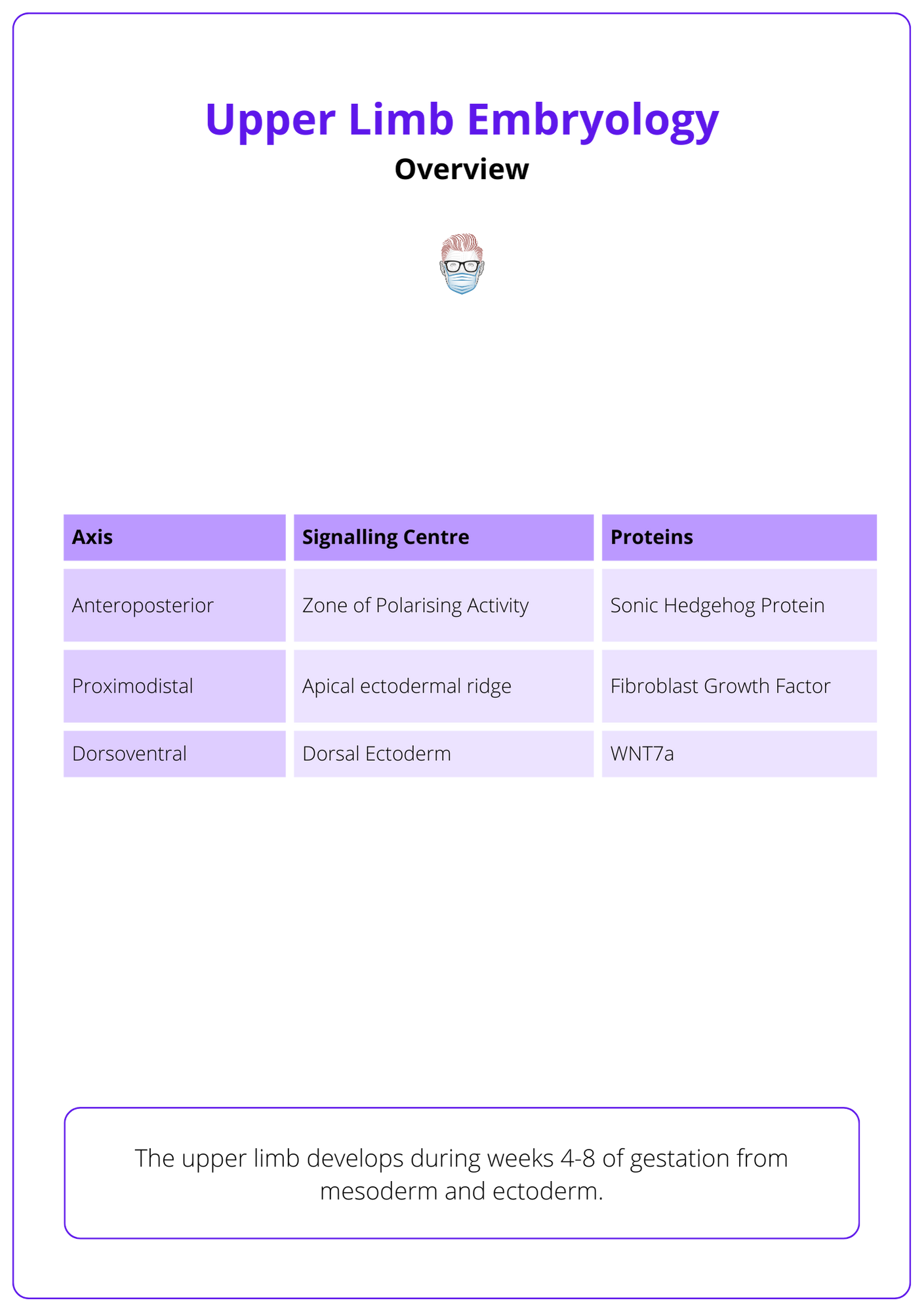
The upper limb develops during weeks 4-8 of gestation from mesoderm and ectoderm. More specifically,
- 3 axis: proximodistal, anteroposterior, dorsoventral
- 3 centres: apical ectodermal ridge, zone of polarizing activity, and dorsal ectoderm.
- 3 proteins: fibroblast growth centre, sonic hedgehog protein, WNT7a.
These critical elements of embryological development are illustrated below.
Developmental Milestones
These are general guidelines for the development milestones of limb embryogenesis.
The upper limb differentiates into a 'human-appearing' limb during 4-8 weeks of gestation. Here are some general timelines to consider:
- ~ Day 26: Limb bud is present.
- ~ Day 30: Blood vessels.
- ~ Day 33: Paddle-shaped hand.
- ~ Day 36: Nerves appear and myofibroblasts become muscle.
- ~ Day 42: Webbed-shaped hand as digital rays become present.
- ~ Day 45: muscles, ligmanets and carpals appear.
- ~ Day 48: Palms move anteriorly and elbows flex.
- ~ Day 50: Digits separate.
- ~ Day 56: Digits ossify secondary to apoptosis.
- ~ Day 58: Human limb.
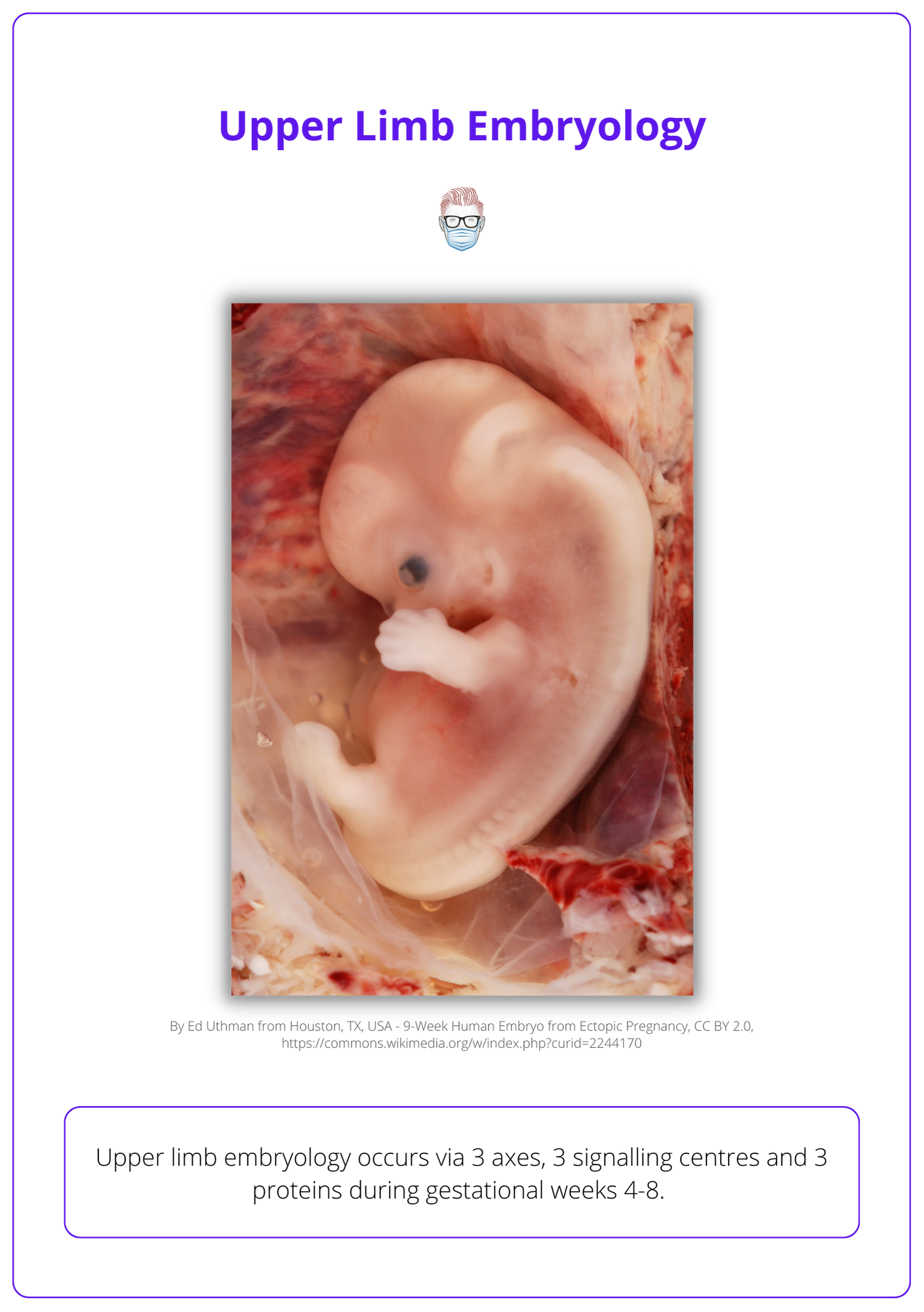
The below image contains a 9-week human embryo and you can observe the upper limb development.
Formation of a Blood Supply
In the 4th week, the limb bud is supplied by a capillary network. This coalesces into a main stem artery that drains into a marginal vein. These structures then allow for the following to occur:
- Subclavian-axillary brachial arterial axis is formed.
- Basilic-axillary-subclavian venous axis is formed.
The brachial artery branches into interosseous and median arteries. The median artery is the main blood supply to the hand, which is then replaced by the ulnar and radial arteries around day 44. The median artery eventually supplies blood to the median nerve.
- Ulnar and radial arteries replaced median artery (now supplies median nerve).
Mechanisms of Development
Upper limb embryology occurs via 3 axes, 3 signalling centres and 3 proteins.
The three axes of of limb development are proximodistal, anteroposterior, and dorsoventral.
Proximodistal Axis
- Role: "distalises" the limb by differentiating limb from the shoulder to finger.
- Signalling centre: Apical Ectodermal Ridge (AER).
- Proteins: Fibroblast growth factors (FGFR 2, 4, 8) influence SHH expression.
Anteroposterior
- Role: "ulnarises" the limb by controlling ulnar and radial growth.
- Signalling centre: Zone of Polarising Activity (ZPA).
- Proteins: Sonic Hedgehog (SHH), which also influences FGFR expression.
Dorsoventral Axis
- Role: "dorsalises" the limb by differentiating a dorsal and palmar surface.
- Signalling centre: Dorsal ectoderm.
- Proteins: WNT7A, which activates LMX1.
Importantly, AER and ZPA are "symbitoic" which results in many conditions affected by the anterioposterior and proximodistal axes:
- SHH signal is required to maintain AER integrity.
- FGFs from the AER are required to express SHH.
There is more information in this webinar below, by Pulvertarft:
Pulvertaft Webinar
Conclusion
1. Basics of Upper Limb Development: You've learned about the embryological processes that occur from weeks 4 to 8 of gestation, involving the mesoderm and ectoderm, leading to the formation of the upper limb.
2. Developmental Milestones: You now know the key developmental milestones, from the initial appearance of the limb bud to the differentiation into a human-appearing limb.
3. Mechanisms of Limb Development: You've explored the three axes of development (proximodistal, anteroposterior, dorsoventral), the three signaling zones (AER, ZPA, dorsal ectoderm), and the three critical proteins (FGFs, SHH, WNT7a) in the process of limb development.
4. Clinical Applications: You have gained a foundational understanding of recognizing developmental anomalies and their potential clinical implications.
