


In this week's edition
- ✍️ Letter from P'Fella
Information has a shelf life - 🤓 The Sunday Quiz
Get ready for week six! - 🖼️ Image of the Week
The perineal body and pelvic floor support - 🚑 Technique Tip
Bilateral gracilis flaps for perineal reconstruction - 📖 What Does the Evidence Say?
Does flap reconstruction reduce wound complications after APR? - 🔥 Articles of the Week
Extended gracilis, VRAM, and perforator flaps for perineal recon. - 💕 Feedback
Suggest ideas & give feedback!
A Letter from P'Fella
Information Has a Shelf Life
It has just been written, referenced, reviewed, illustrated and checked. Then we press publish and move on to the next one.
But the subject doesn’t stop moving with us.
The guidelines update. A new paper challenges something we previously accepted. An operation evolves. Someone notices that an illustration could be clearer. Occasionally, it’s something much smaller: a dose, classification, or recommendation that was correct when the article was written but no longer reflects current practice.
This has always made it clear what it means to run a medical education platform.
Publishing something is relatively easy to monitor. We can count how many articles we have written this month or how many new topics we have added to the Library. What is much harder to see is the work happening to the hundreds of articles that are already there. And arguably, that is a bigger responsibility than writing new articles.
The most concerning outdated information isn’t usually an article that is obviously ten years behind. You can recognise that. It’s an otherwise excellent article with one important paragraph that has become outdated. Everything around it still looks credible, so there is very little reason for the reader to question it.
In the past couple of years, we’ve been putting more thought into what happens after we publish.
Our departments and On-Call Teams are part of that. Rather than treating review as something that happens once before publication, we want people working within each area to keep coming back to the material: reviewing articles, flagging changes in practice, improving illustrations, and telling us when something needs another look.
That's why we’re also working through older parts of the Library systematically. Some articles need a small update. Others need new evidence or illustrations. And occasionally, the right decision is to rebuild the article altogether.
Readers have a role in this too. Some of the most useful corrections we receive start with a simple email saying, “I don’t think this is quite right.” I hope people continue sending those. An educational resource should be able to withstand being questioned, and it should get better when somebody spots something we’ve missed.
There’s a broader lesson here that applies well beyond P’Fella.
When you read something clinical, don’t only ask whether the source is trustworthy. Ask when the information was last challenged.
A perfectly referenced article can still age. A familiar technique can change. And something you learnt for an exam five years ago may deserve another look before you use it to make a decision today.
For us, that means success can’t just be measured by how much we publish. It has to include how well we look after what we’ve already put into the world.
Pressing publish shouldn’t be the end of an article. It's only the start of its maintenance.
P’Fella ❤️
The Sunday Quiz
Get Ready for Week Six!
Each week, we'll put your plastic surgery knowledge to the test with a new question designed to challenge you and keep you thinking. Join in every Sunday, keep your wits about you, and compete for the top spot. The winner at the end of eight rounds will receive 6 months of Plastics Pro, on us.
Image of the Week
The Perineal Body and Pelvic Floor Support
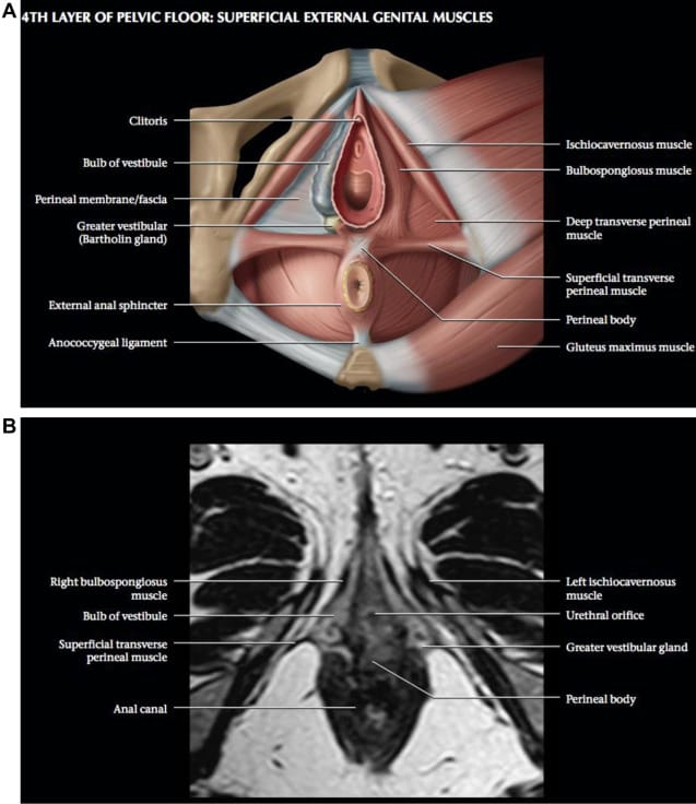
This week's image includes an anatomical illustration with an axial MRI, showing the relationship between the bulbospongiosus, ischiocavernosus, transverse perineal muscles, and external anal sphincter. The perineal body sits between the vaginal opening and anus, where several of these muscles converge to provide structural support.
For perineal reconstruction, restoring skin coverage alone may not be enough. Understanding these muscular attachments is essential when reconstructing defects following trauma, tumour excision, or abdominoperineal resection, where loss of supporting tissues can compromise pelvic floor integrity.
Technique Tip
Bilateral Gracilis Flaps for Perineal Reconstruction
The gracilis muscle flap is a useful option for perineal reconstruction following abdominoperineal excision (APE), particularly in patients with large pelvic floor defects or previously irradiated tissues. Bilateral gracilis flaps provide vascularised tissue to fill the pelvic dead space and reinforce the pelvic floor, with the aim of reducing wound complications and supporting healing.
This video demonstrates pelvic floor reconstruction using bilateral gracilis flaps following APE for rectal cancer.
What Does the Evidence Say?
Does Flap Reconstruction Reduce Wound Complications After APR?
More recent randomised evidence is less definitive. In the BIOPEX-2 trial, a gluteal turnover flap did not improve uncomplicated wound healing at 30 days compared with primary closure (55% vs 60%). Perineal infection, wound dehiscence requiring intervention, and perineal hernia were also not significantly reduced. The clearest advantage was in deeper infection: presacral abscess occurred in 9% after flap closure versus 22% after primary closure, while percutaneous drainage was required in 1% versus 8%. Importantly, this trial evaluated a gluteal turnover flap, not VRAM, and excluded the most extensive pelvic resections.
The benefit appears strongest in previously irradiated patients. In a cohort of 329 patients undergoing APR after neoadjuvant radiotherapy, delayed healing at 3 months occurred in 31.5% after direct closure versus 10.4% after VRAM reconstruction; pelvic abscess occurred in 26.9% versus 10.1%. These results support bringing well-vascularised tissue into an irradiated defect, but they remain non-randomised and vulnerable to selection bias. Overall, flap reconstruction does not consistently prevent superficial wound breakdown or perineal hernia after every APR, but it can reduce pelvic abscesses and may improve healing in irradiated or otherwise high-risk defects.
(Devulapalli, 2016); (Kreisel, 2025); (Spasojevic, 2018); (Buscail, 2021); (Copeland-Halperin, 2020)
Articles of the Week
3 Interesting Articles with One-Sentence Summaries
Extended gracilis dissection increases the flap’s arc of rotation, allowing greater coverage of complex perineal and groin wounds, with 100% successful reconstructions in 19 high-risk patients and only one reported complication.
VRAM reconstruction had a 27% perineal complication rate, but complete flap loss was uncommon at just 1.8%, highlighting its reliability despite considerable postoperative morbidity.
Perineal perforator-based island flaps preserve muscle and major donor vessels, with no flap loss or donor-site complications in 11 patients, offering a promising alternative to conventional flaps with minimal donor-site morbidity.
