


In this week's edition
- ✍️ Letter from P'Fella
Clinical illustrations that actually teach - 🤓 The Sunday Quiz
Keep your wits about you for week two! - 🖼️ Image of the Week
Does facial fat move with gravity? - 🚑 Technique Tip
Retaining ligament release in deep plane facelift - 🔦 Spotlight
ST3 portfolio and interview guide - 📖 What Does the Evidence Say?
What actually causes facial ageing? - 🔥 Articles of the Week
Facial assessment before fillers & facial ageing. - 💕 Feedback
Suggest ideas & give feedback!
A Letter from P'Fella
Clinical Illustrations That Actually Teach
Images in surgical education are harder to get right than they first appear. Published figures come with copyright and licensing restrictions. Clinical photographs need consent, careful anonymisation, and proper documentation. Even when a usable image exists, it may show the anatomy from the wrong angle, include too much detail, or fail to explain the particular point being made in the article.
We decided to build a more consistent system of our own.
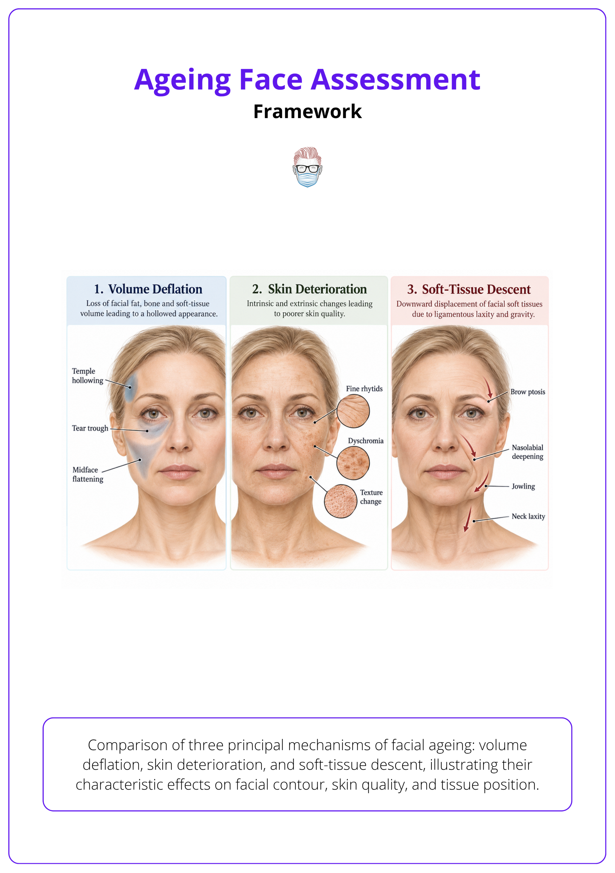
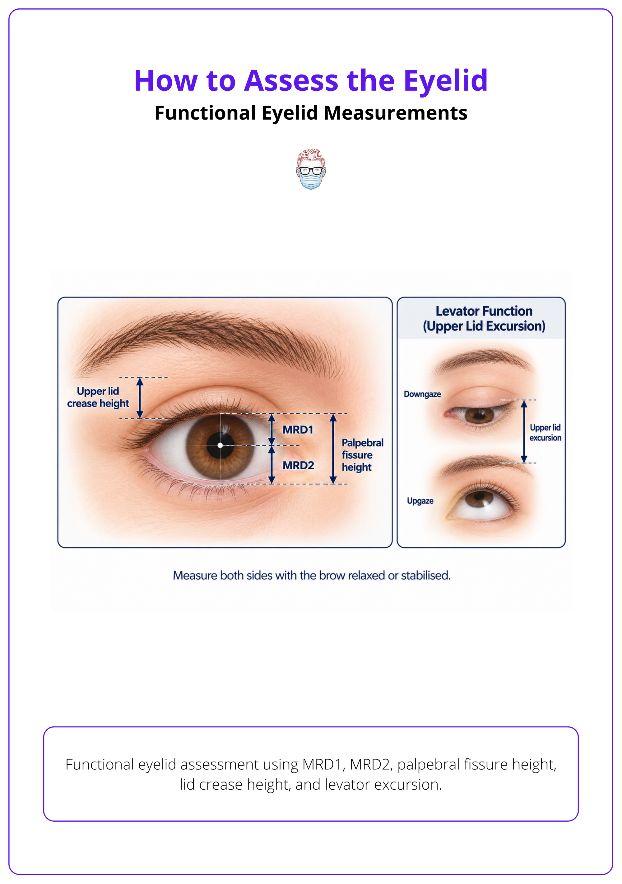
Clinical Illustrations
The examples here show where we’ve reached. One maps the key neurovascular structures around the jawline and chin. Another separates the main components of facial ageing. The eyelid illustration brings several functional measurements together in one view. Each was made for a specific section of an article, with the labels, perspective, and level of detail chosen around what the reader needs to understand at that moment.
That last part has become the most important part of the process. A clinical illustration should have a clear job. It might orientate you to the anatomy before an assessment, show the relationship between structures, or make a measurement easier to remember. If it cannot improve the explanation, it probably does not need to be there.
We’re now using this approach in several newly published articles:
We’re confident in the direction and ready to begin applying it more widely across the platform. Before we do, I’d like you to see the illustrations where they belong: inside the articles, alongside the ideas they were made to explain.
Have a read and send us your honest thoughts. Tell us which illustrations made the subject clearer, and where a label, detail, or teaching point could still be sharpened. That feedback will help us make a strong process even more consistent as we bring it into the rest of the curriculum.
P’Fella ❤️
The Sunday Quiz
Keep Your Wits About You for Week Two
Each week, we'll put your plastic surgery knowledge to the test with a new question designed to challenge you and keep you thinking. Join in every Sunday, keep your wits about you, and compete for the top spot. The winner at the end of eight rounds will receive 6 months of Plastics Pro, on us.
Image of the Week
Does Facial Fat Move with Gravity?
Facial ageing isn't just about losing volume. Where fat sits within the face may be just as important as how much is present.
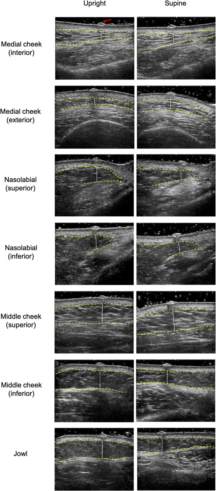
These ultrasound images compare superficial facial fat thickness in upright and supine positions. When lying down, the medial and upper middle cheek become thicker, while the nasolabial, lower middle cheek, and jowl regions become thinner. The findings support the theory that superficial fat can redistribute within its compartments in response to changes in position.
The surgical implication is this: what appears to be facial volume loss may sometimes reflect fat redistribution rather than true depletion. Recognising this distinction could help surgeons decide when to restore volume and when to reposition existing tissue.
Technique Tip
Retaining Ligament Release in Deep Plane Facelift
The release of facial retaining ligaments is a key step in deep plane facelift surgery, allowing mobilisation and repositioning of the superficial musculoaponeurotic system (SMAS) and overlying soft tissues. Adequate release reduces tethering and permits tissue repositioning with less reliance on skin tension, while careful dissection is essential to protect the facial nerve.
This operative video demonstrates deep plane dissection and the release of retaining ligaments, highlighting the anatomical relationships between the zygomaticus major, masseter, and adjacent soft tissues. The technique illustrates how ligament release facilitates mobilisation and resuspension of the midface during facial rejuvenation surgery.
Spotlight
ST3 Portfolio and Interview Guide
We've put together an updated 3-part ST3 Guide Series, authored by Carissa Jacobs, covering everything from eligibility and portfolio scoring to interview preparation, clinical scenarios, and communication.
The series includes practical preparation tips, common pitfalls, and advice to help you prepare for the standard expected of an incoming ST3 registrar.
Plastic Surgery ST3 Guide
- Eligibility & Portfolio Scoring: Eligibility, evidence, and portfolio scoring.
- Interview Format & Preparation: How the interview works and what is expected.
- Clinical Scenarios & Communication: Clinical reasoning, prioritisation, consent, and calling a senior.
What Does the Evidence Say?
What Actually Causes Facial Ageing?
Facial fat does not age as one continuous layer either. Anatomical studies show that it is divided into discrete compartments, while CT and MRI studies demonstrate that these compartments behave differently: some migrate inferiorly, some lose volume, and others may retain or gain volume. Deep medial cheek fat loss, lower-pole redistribution, and selective upper-cheek fat enlargement have all been described, showing why hollowing and fullness can appear beside one another in the same ageing face.
Soft-tissue descent still contributes, but it is not uniform across the face. Longitudinal photographic analysis found remarkably little downward movement of the lid-cheek junction and upper midface over several decades, challenging the idea that gravity alone explains these changes.
The practical takeaway is that facial ageing results from an interaction between skeletal remodelling, compartment-specific fat change, and variable soft-tissue descent. Assessment should therefore identify which layer is driving each visible feature rather than treating every face as simply sagging or volume-deficient.
(Shaw Jr, 2007); (Rohrich, 2007); (Gierloff, 2012); (Gosain, 2005); (Lambros, 2007)
Articles of the Week
3 Interesting Articles with One-Sentence Summaries
A clinical review on 8 parameters for facial assessment, including facial proportions, tissue depth and quality, symmetry, dynamic movement, ethnicity and gender ideals, to guide individualised filler treatment.
Deep facial fat compartments tend to lose volume, while superficial compartments may enlarge or descend, contributing to tear troughs, nasolabial folds, and jowls, and supporting a targeted approach to volume restoration rather than lifting alone.
Bone resorption occurs predictably in the orbital rims, maxilla, pyriform aperture, and prejowl region, contributing to tear troughs, nasolabial folds, and jowls, which supports restoring skeletal support being as important as soft-tissue repositioning in facial rejuvenation.
