


In this week's edition
- ✍️ Letter from P'Fella
Teaching plastic surgery is becoming more visual - 🖼️ Image of the Week
Retrograde venous flap for dorsal finger reconstruction - 🚑 Technique Tip
Zone II flexor tendon repair - 🎧 How I Operate
Designing the perfect PIA flap with Professor Claudio Angrigiani - 📖 What Does the Evidence Say?
Dupuytren’s treatment: Recovery vs durability - 🔥 Articles of the Week
Hand vascular anatomy, and metacarpal & phalangeal fractures - 💕 Feedback
Suggest ideas & give feedback!
A Letter from P'Fella
Teaching Plastic Surgery Is Becoming More Visual
You can describe an anatomical relationship in a paragraph, but sometimes a single illustration communicates the idea instantly. That's always been one of the challenges in surgical education. Not every concept has a good image to accompany it. Even when one exists, using it isn't always straightforward.
Clinical photographs require consent and meticulous attention to patient confidentiality, while illustrations and published figures are often protected by copyright. By the time you've navigated permissions, documentation, and licensing, creating educational content can become a project in itself.
Over the last few months, we've been building a new image generation platform by The Plastics Fella. It's something we've experimented with for a while, and the technology has improved dramatically over the past three months. We're now at the point where it can generate clinically accurate anatomy and patient illustrations that are useful for teaching and learning.
The actual challenge isn't creating the images. It's making them educational. We're currently developing tools that allow us to label structures, highlight anatomical landmarks, compare clinical photographs, and annotate illustrations in a way that's accurate and easy to understand. Instead of simply looking at an image, you'll be able to create it, test yourself, and understand how a clinical concept actually works.
I'm excited to share a few early examples of what we've been working on. We currently have a range of simplified anatomical illustrations to clinical photographs paired with interactive labels. They're still works in progress, but this is making impressive progress and I'm hoping to share it with you soon.
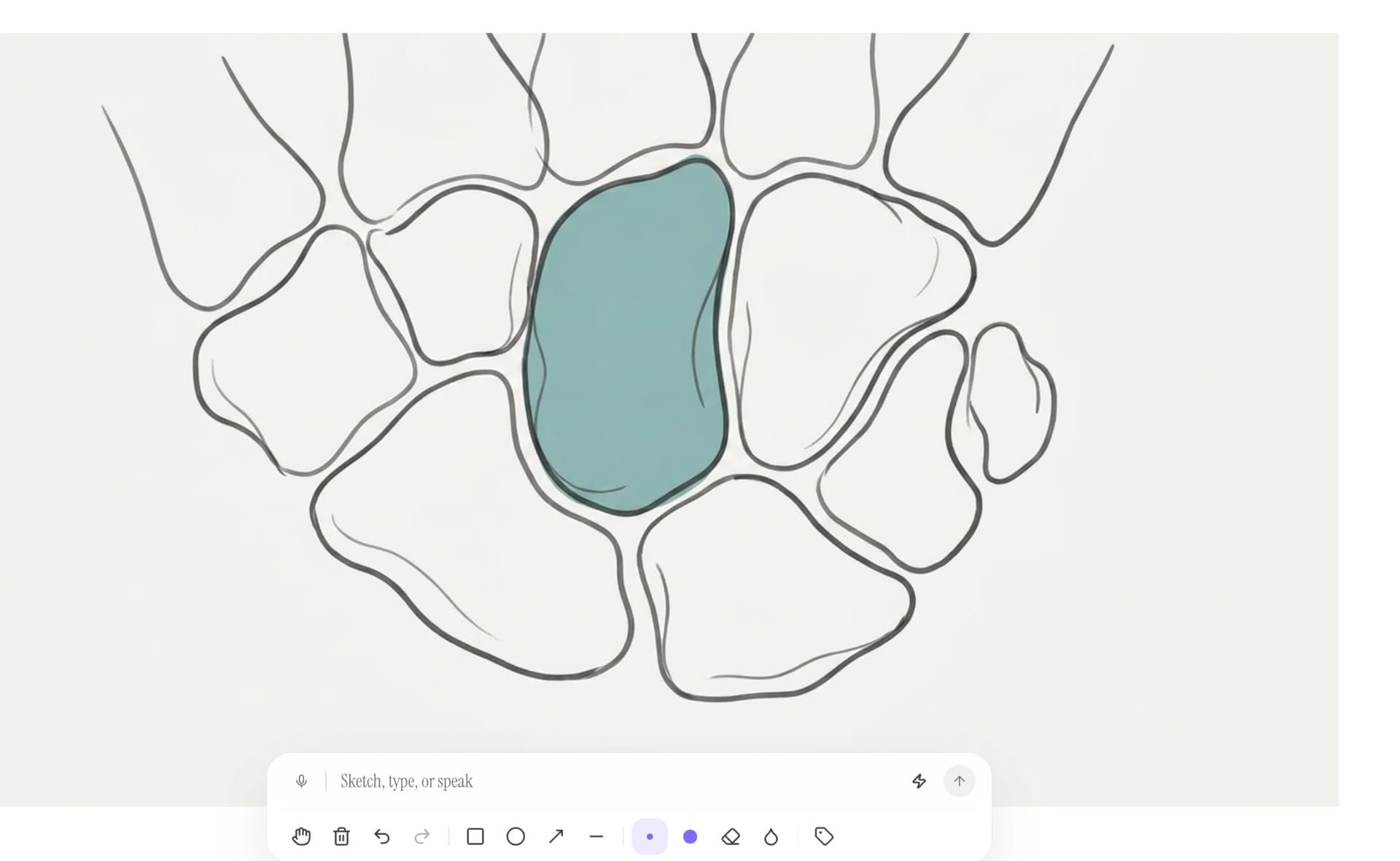
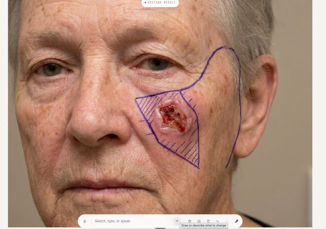
Visual Tool for Plastic Surgery Learning
Our goal has always been the same: make plastic surgery easier to learn. If better tools help someone understand anatomy a little faster or remember an operation for longer, then they're worth building.
P’Fella ❤️
Image of the Week
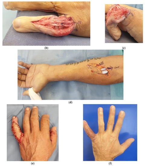
Retrograde Venous Flap for Dorsal Finger Reconstruction
This case shows a complex dorsal finger defect following trauma, with tendon adhesions and joint pathology requiring tenoarthrolysis and joint replacement. The key reconstructive step is the use of a retrograde free venous flap harvested from the forearm.
Unlike conventional arterial flaps, this technique uses a venous conduit that is arterialised, with the vein anastomosed to a digital artery to provide inflow. This allows thin, pliable tissue coverage, which is ideal for the dorsum of the finger while preserving contour and enabling tendon gliding.
The sequence highlights the principle clearly: restore function first (joint and tendon), then provide appropriate soft tissue coverage that won’t compromise movement.
Technique Tip
Zone II Flexor Tendon Repair
Zone II remains one of the most technically demanding regions for flexor tendon repair. Success depends on meticulous tendon handling, a strong core suture construct, preservation of the pulley system where possible, and a repair that permits early controlled mobilisation without excessive bulk or gapping.
This operative video demonstrates a Zone II flexor tendon repair, highlighting tendon retrieval, core and epitendinous suturing, and techniques to achieve a smooth repair that glides freely within the flexor sheath.
How I Operate
Designing the Perfect PIA Flap with Professor Claudio Angrigiani
Professor Claudio Angrigiani challenges the way we describe “fasciocutaneous” flaps. His point is simple: a flap is skin and subcutaneous tissue transferred while preserving its vascularisation. The fascia may help with dissection or handling, but it is not necessarily what carries the blood supply. So, include the fascia if it helps the operation, but don’t confuse fascia with the flap.
What Does the Evidence Say?
Dupuytren’s Treatment: Recovery vs Durability
Long-term evidence favours limited fasciectomy for durability. In a randomised comparison, recurrence at five years was 84.9% after needle fasciotomy versus 20.9% after limited fasciectomy, although many patients still preferred the minimally invasive procedure because of its quicker recovery and lower treatment burden (van Rijssen et al., 2012). More recent randomised evidence found that collagenase produced worse patient-reported hand outcomes than limited fasciectomy at one and two years and was associated with more recurrent contractures requiring reintervention (8% vs 1.7%). However, moderate-to-severe complications were less frequent after collagenase (2% vs 5%).
The practical takeaway is that there is no universally superior treatment. Minimally invasive procedures may suit older patients, those seeking rapid recovery, or those willing to accept repeat treatment. Limited fasciectomy offers a more durable correction and appears particularly valuable for more advanced contractures, but this must be balanced against its greater surgical and recovery burden. Treatment choice should therefore be based not only on the initial correction, but on how the patient values recovery time, procedural risk, and the likelihood of recurrence.
(van Rijssen, 2012); (Dias, 2024); (Hurst, 2009); (Rodrigues, 2015)
Articles of the Week
3 Interesting Articles with One-Sentence Summaries
This review maps the radial, ulnar, and interosseous arterial systems and their perforators, showing how their interconnected arches and digital branches underpin the design and reliability of local and regional hand flaps.
Stable fractures can be managed nonoperatively, but when fixation is required, outcomes depend less on the hardware and more on preserving soft tissue and restoring precise rotational alignment to maintain hand function.
Metacarpal fracture management should prioritise function over radiographic perfection, as operative fixation can improve alignment but risks stiffness, making careful patient and fracture selection critical.
