


In this week's edition
- ✍️ Letter from P'Fella
Our first specialist edition is almost here - 🖼️ Image of the Week
Reading ICG lymphography - 🚑 Technique Tip
ICG-guided lymphatic mapping - 📖 What Does the Evidence Say?
Reverse lymphatic mapping in VLNT - 🔥 Articles of the Week
Lymphoscintigraphy & lymphedema patterns for LVA. - 💕 Feedback
Suggest ideas & give feedback!
A Letter from P'Fella
Our First Specialist Edition Is Almost Here
And, the first edition is almost ready!
We're starting with Aesthetics, and putting this together has helped us work out what we actually want these monthly editions to be. We didn't want just an email filled with links to papers or a long overview of a huge topic. There are already plenty of places to find information. We wanted to build something that you could read in 10-15 minutes and come away thinking a little differently about a problem you might actually see in clinic.
The first edition is a good example. We're focusing entirely on periorbital assessment. First, we bring up a clinical question with context. From there, we work through the findings that might change your plan, how to do the assessment, the common misconceptions you might fall into, and eventually the decision itself

With these specialist editions, we planned on going in the direction of recreating a clinical scenario you will likely come across. Each month, we want to take one useful clinical problem and properly work through it, with input from P'Fella's specific department. Our current structure is: The case, assessment, thinking behind the decision, what to avoid, and evidence worth knowing.
Aesthetics is where we're starting, and if this format works, there's plenty of room to take it into the other departments we're building too. Each specialty has those decisions and principles that are difficult to pick up from reading facts alone, and I'd love for these editions to become somewhere we can unpack them properly.
For now, we're doing the finishing touches on Edition 1.
If this specialist aesthetics newsletter is something you're interested in, join the mailing list below, and we'll send you the first edition when it goes live.
Hope you enjoy what we've been building.
P’Fella ❤️
Image of the Week
Reading ICG Lymphography
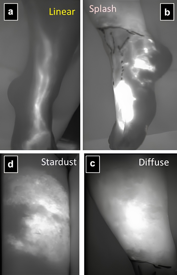
These four Indocyanine green (ICG) lymphography patterns show the progression from normal lymphatic flow to increasingly abnormal dermal backflow.
A linear pattern represents normal collecting lymphatics. With lymphatic dysfunction, this changes to splash, then stardust, and eventually a diffuse pattern as lymphatic damage becomes more severe.
These changes can appear before obvious swelling develops, making ICG lymphography particularly useful for detecting early lymphatic dysfunction. It also provides real-time mapping of superficial lymphatic flow, which can help identify suitable lymphatics for LVA.
Technique Tip
ICG-Guided Lymphatic Mapping
ICG lymphography uses near-infrared fluorescence to provide real-time visualisation of superficial lymphatic drainage. Following injection, movement of ICG through afferent lymphatic vessels can be followed towards the regional nodal basin, allowing lymphatic channels and sentinel nodes to be identified intraoperatively.
In this operative segment, ICG demonstrates defined lymphatic transit towards the axilla, with an afferent lymphatic channel leading to two fluorescent sentinel nodes. The fluorescence overlay allows these structures to be localised relative to the surrounding surgical anatomy during dissection.
What Does the Evidence Say?
Reverse Lymphatic Mapping in VLNT
The original prospective PRS description used differential tracers to identify limb-draining versus trunk-draining nodes during VLNT harvest. Subsequent work refined the technique using indocyanine green (ICG) and blue dye, showing that ICG mapping identified the same key lower-extremity draining nodes as technetium-99 mapping in all 39 patients studied, with no donor-site lymphedema reported. More recent prospective JPRAS data are similarly reassuring: among 51 patients undergoing groin VLNT with reverse lymphatic mapping, none developed a lower-extremity volume difference greater than 10% at 12 months.
The practical takeaway is that reverse lymphatic mapping should be viewed as a risk-reduction strategy, not a guarantee. The evidence is still based mainly on prospective cohort studies rather than randomized trials, but it consistently supports the same principle: if lymph nodes are being harvested from a basin that also drains an uninvolved limb, identifying and preserving those limb-draining nodes is one of the most defensible ways to reduce the risk of creating a new lymphedema problem while treating another.
(Chang, 2022); (Schaverien, 2018); (Pajula, 2024); (Pons, 2021); (Dayan, 2015)
Articles of the Week
3 Interesting Articles with One-Sentence Summaries
Quantitative lymphoscintigraphy detected abnormal lymphatic function in all 308 affected limbs in one cited series, including mild cases missed by qualitative imaging, highlighting its value when early lymphedema is clinically subtle.
Lymphoscintigraphy distinguished lymphedema from other causes of limb swelling, with 80% sensitivity and 94% specificity, and excluded lymphedema in approximately one-third of referred patients.
Among 142 limbs classified by lymphoscintigraphy, type III - dermal backflow involving both the thigh and leg - was identified as the most favourable pattern for lymphaticovenous anastomosis.
